

Trouble-makers in cytologic interpretation of the uterine cervix
- PMID: 37194148
- PMCID: PMC10209661
- DOI: 10.4132/jptm.2023.04.25
Trouble-makers in cytologic interpretation of the uterine cervix
Abstract
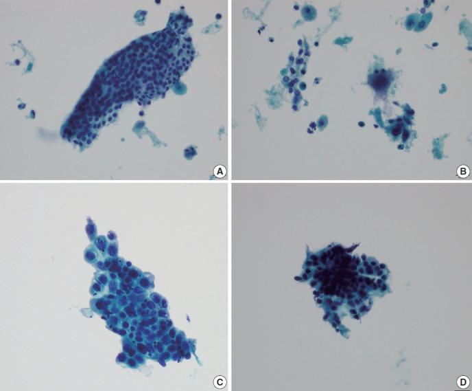
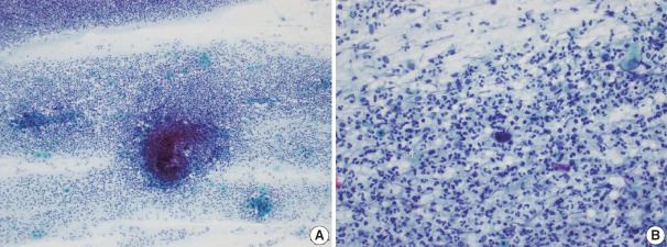
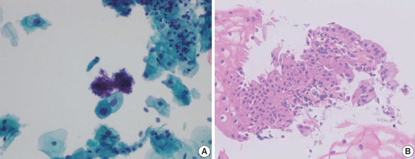
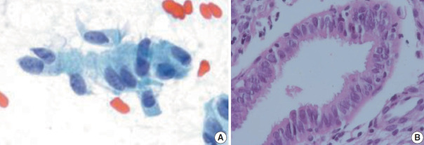
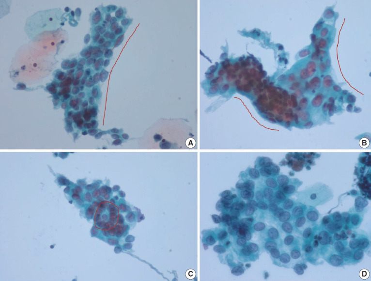
The development and standardization of cytologic screening of the uterine cervix has dramatically decreased the prevalence of squamous cell carcinoma of the uterine cervix. Advances in the understanding of biology of human papillomavirus have contributed to upgrading the histologic diagnosis of the uterine cervix; however, cytologic screening that should triage those that need further management still poses several difficulties in interpretation. Cytologic features of high grade intraepithelial squamous lesion (HSIL) mimics including atrophy, immature metaplasia, and transitional metaplasia, and glandular lesion masquerades including tubal metaplasia and HSIL with glandular involvement are described with accentuation mainly on the differential points. When the cytologic features lie in a gray zone between the differentials, the most important key to the more accurate interpretation is sticking to the very basics of cytology; screening the background and cellular architecture, and then scrutinizing the nuclear and cytoplasmic details.
Keywords: Atrophy; Cytology; Glandular involvement; High grade intraepithelial squamous lesion; Metaplasia; Uterine cervix.
Figures
Similar articles
-
Papillary immature metaplasia of the uterine cervix: a report of 5 cases with an emphasis on the differential diagnosis from reactive squamous metaplasia, high-grade squamous intraepithelial lesion and papillary squamous cell carcinoma.J Korean Med Sci. 2001 Dec;16(6):762-8. doi: 10.3346/jkms.2001.16.6.762. J Korean Med Sci. 2001. PMID: 11748359 Free PMC article.
-
Transitional cell metaplasia of the uterine cervix is related to human papillomavirus: molecular analysis in seven patients with cytohistologic correlation.Cancer. 2002 Aug 25;96(4):250-8. doi: 10.1002/cncr.10722. Cancer. 2002. PMID: 12209668
-
Cytologic correlates of papillary immature metaplasia (immature condyloma) of the cervix.Diagn Cytopathol. 1998 Jun;18(6):416-21. doi: 10.1002/(sici)1097-0339(199806)18:6<416::aid-dc6>3.0.co;2-6. Diagn Cytopathol. 1998. PMID: 9626513
-
Practical issues related to uterine pathology: in situ and invasive cervical glandular lesions and their benign mimics: emphasis on cytology-histology correlation and interpretive pitfalls.Mod Pathol. 2016 Jan;29 Suppl 1:S1-11. doi: 10.1038/modpathol.2015.138. Mod Pathol. 2016. PMID: 26715169 Review.
-
Colposcopy, cervicography, speculoscopy and endoscopy. International Academy of Cytology Task Force summary. Diagnostic Cytology Towards the 21st Century: An International Expert Conference and Tutorial.Acta Cytol. 1998 Jan-Feb;42(1):33-49. doi: 10.1159/000331533. Acta Cytol. 1998. PMID: 9479322 Review.
References
-
- Acs G, Gupta PK, Baloch ZW. Glandular and squamous atypia and intraepithelial lesions in atrophic cervicovaginal smears: one institution’s experience. Acta Cytol. 2000;44:611–7. - PubMed
-
- Kaminski PF, Sorosky JI, Wheelock JB, Stevens CW., Jr The significance of atypical cervical cytology in an older population. Obstet Gynecol. 1989;73:13–5. - PubMed
-
- Waddell CA. The influence of the cervix on smear quality. I: Atrophy. An audit of cervical smears taken post-colposcopic management of intraepithelial neoplasia. Cytopathology. 1997;8:274–81. - PubMed
-
- Mokhtar GA, Delatour NL, Assiri AH, Gilliatt MA, Senterman M, Islam S. Atypical squamous cells, cannot exclude high-grade squamous intraepithelial lesion: cytohistologic correlation study with diagnostic pitfalls. Acta Cytol. 2008;52:169–77. - PubMed
Publication types
LinkOut - more resources
Full Text Sources
