. 2023 Sep;43(9):1503-1518.
doi: 10.1177/0271678X231175190. Epub 2023 May 17.

Neutrophil-derived cathelicidin promotes cerebral angiogenesis after ischemic stroke

Affiliations

Neutrophil-derived cathelicidin promotes cerebral angiogenesis after ischemic stroke

Wanqing Xie et al. J Cereb Blood Flow Metab. 2023 Sep.

Abstract

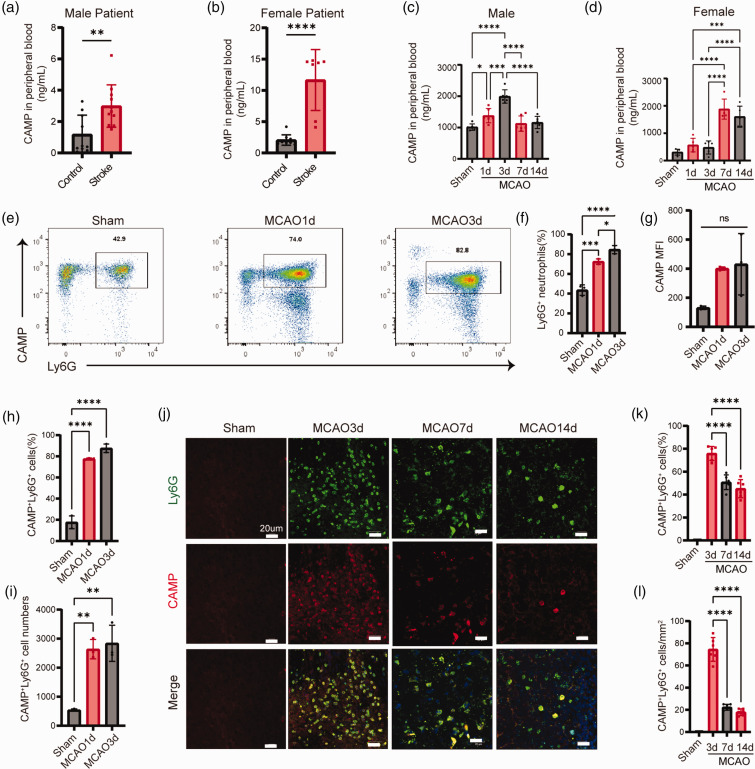
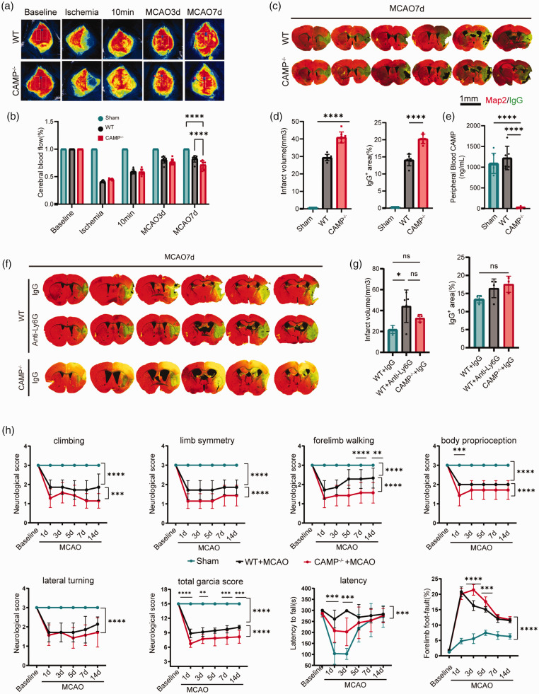
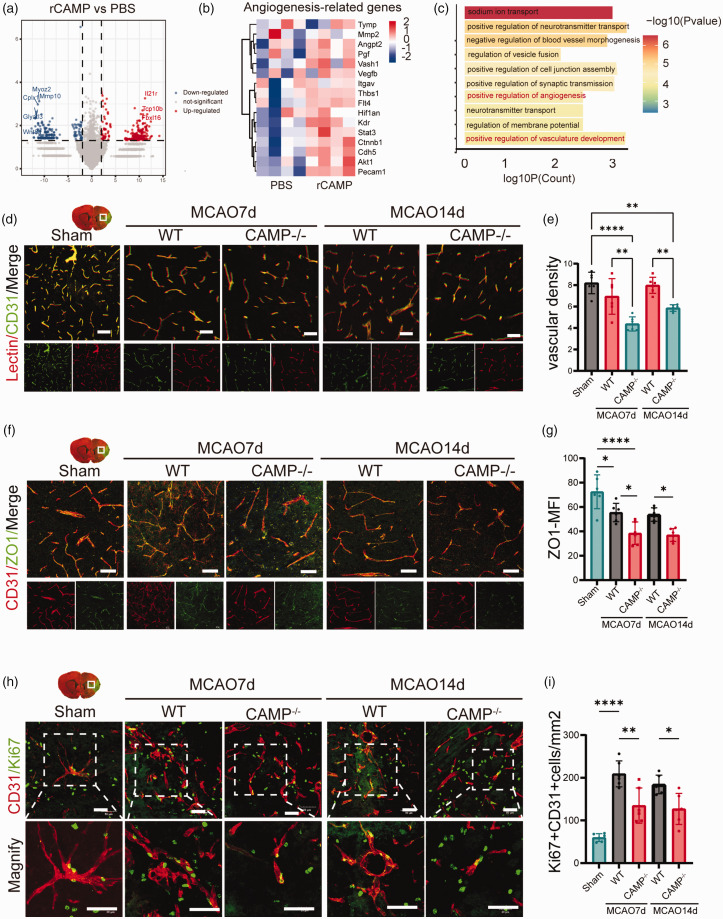
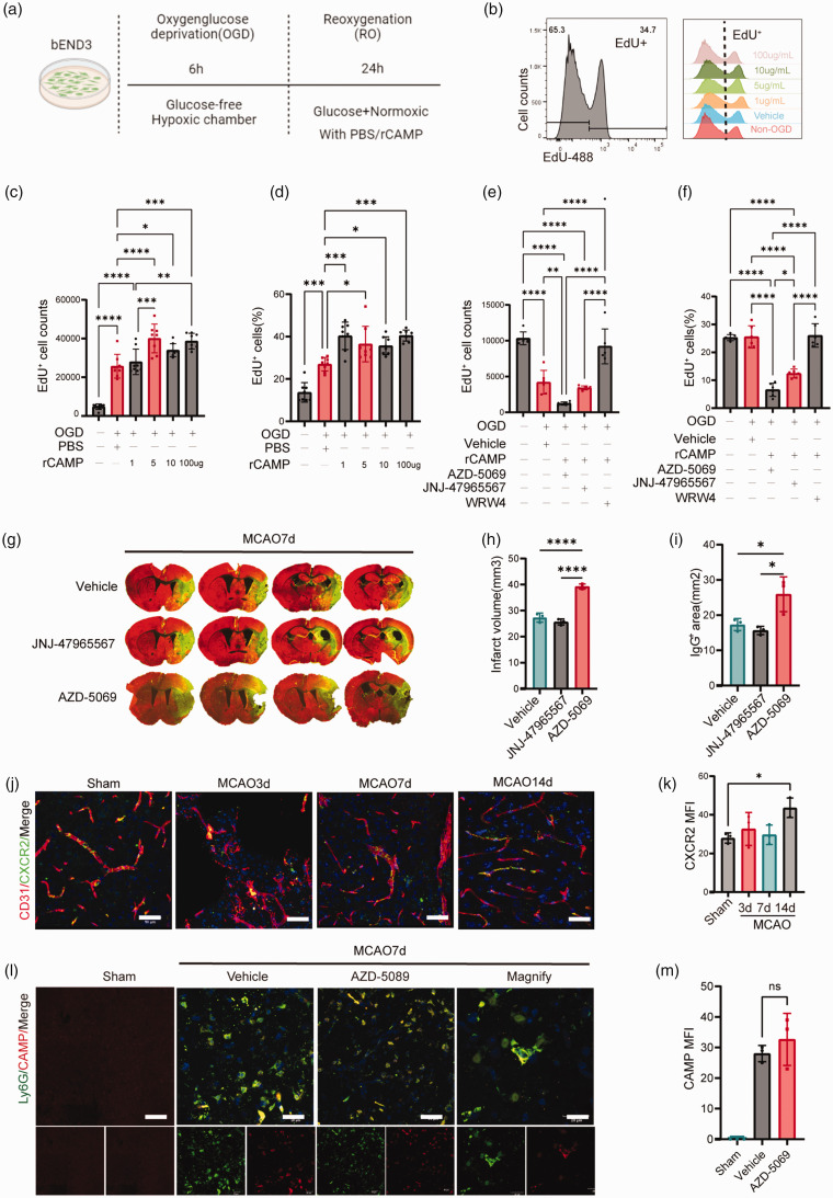
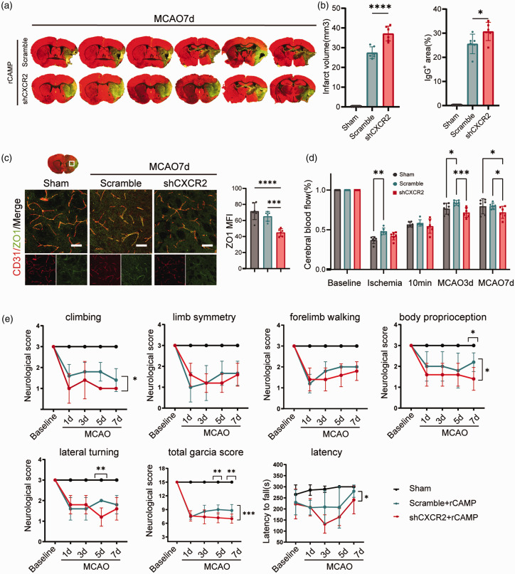
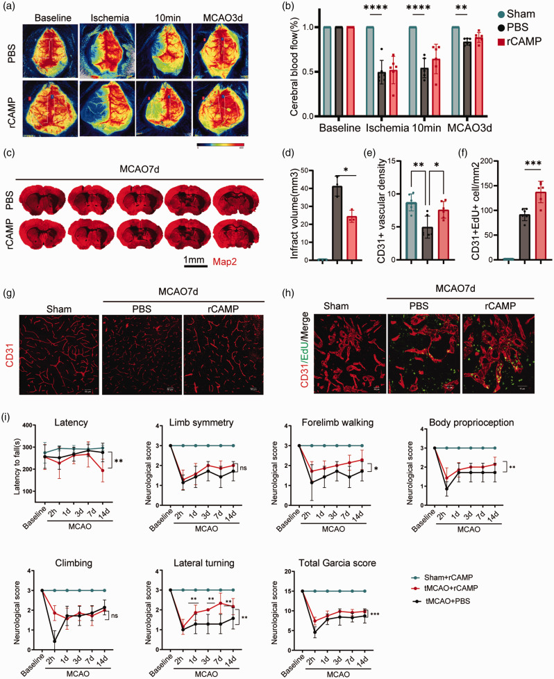
Neutrophils play critical roles in the evolving of brain injuries following ischemic stroke. However, how they impact the brain repair in the late phase after stroke remain uncertain. Using a prospective clinical stroke patient cohort, we found significantly increased cathelicidin antimicrobial peptide (CAMP) in the peripheral blood of stroke patients compared to that of healthy controls. While in the mouse stroke model, CAMP was present in the peripheral blood, brain ischemic core and significantly increased at day 1, 3, 7, 14 after middle cerebral artery occlusion (MCAO). CAMP-/- mice exhibited significantly increased infarct volume, exacerbated neurological outcome, reduced cerebral endothelial cell proliferation and vascular density at 7 and 14 days after MCAO. Using bEND3 cells subjected to oxygen-glucose deprivation (OGD), we found significantly increased angiogenesis-related gene expression with the treatment of recombinant CAMP peptide (rCAMP) after reoxygenation. Intracerebroventricular injection (ICV) of AZD-5069, the antagonist of CAMP receptor CXCR2, or knockdown of CXCR2 by shCXCR2 recombinant adeno-associated virus (rAAV) impeded angiogenesis and neurological recovery after MCAO. Administration of rCAMP promoted endothelial proliferation and angiogenesis and attenuated neurological deficits 14 days after MCAO. In conclusion, neutrophil derived CAMP represents an important mediator that could promote post-stroke angiogenesis and neurological recovery in the late phase after stroke.

Keywords: Ischemic stroke; angiogenesis; cathelicidin; cerebral endothelial cell; neutrophil.


Conflict of interest statement

The author(s) declared no potential conflicts of interest with respect to the research, authorship, and/or publication of this article.

Figures

Figure 1.
Figure 1.
CAMP is upregulated in circulating neutrophils and ischemic brain. (a–b) Quantification of CAMP in the plasma of male (a) and female (b) stroke patients within 3 days by ELISA (n = 17-23 per group, unpaired Student’s t-test). (c–d) Quantification of CAMP in the plasma of MCAO male mice (c) and female mice (d) at 1, 3, 7 and 14 days after MCAO (n = 6 per group, one-way ANOVA with Bonferroni multiple comparisons test). (e) Representative flow cytometry plots for CAMP+ Ly6G+ neutrophils in the peripheral blood of MCAO mice at 1 and 3 days after MCAO. (f–i) Quantification of CAMP+ Ly6G+ neutrophils cell numbers and percentage, total neutrophils prcentage and CAMP MFI in the peripheral blood at 1 and 3 days after MCAO (n = 3 per group, one-way ANOVA with Bonferroni multiple comparisons test). (j) Representative images showed expression of CAMP in neutrophils inflitrated in the infarct core at indicated timepoints after MCAO. The representative pictures of three individual mouse samples per group were shown. Scale bar, 20μm and (k–l) Quantification of CAMP+ Ly6G+ neutrophils counts per mm2 and percentage in the infarct core at indicated timepoints after MCAO (n = 6 per group, one-way ANOVA with Bonferroni multiple comparisons test). All data are presented as means ± SD, *P < 0.05, **P < 0.01, ***P < 0.001, ****P < 0.0001, ns, no significance.
Figure 2.
Figure 2.
Neutrophil-derived CAMP deficiency increases infarct volume and impairs neurological recovery after ischemic stroke. (a) Representative images of cerebral blood flow (CBF) of WT and CAMP−/− mice before MCAO (baseline), during ischemia, 10 minutes, 3 days and 7 days after reperfusion. (b) Quantitative measurements of CBF of Sham, WT and CAMP−/− mice at indicated timepoints. Results were expressed as percent change from baseline (n = 8 per group, one-way ANOVA with Bonferroni multiple comparisons test). (c) Representative MAP2 staining of brain infarct and endogenous mouse IgG staining of BBB leakage of WT and CAMP−/− mice 7 days after MCAO. Scale bar, 1 mm. (d) Quantification of infarct volume and endogenous IgG positive area of WT and CAMP−/− mice 7 days after MCAO (n = 8 per group, one-way ANOVA with Bonferroni multiple comparisons test). (e) Quantification of CAMP in the plasma of WT and CAMP−/− male mice 7 days after MCAO (n = 6 per group, one-way ANOVA with Bonferroni multiple comparisons test). (f) Representative MAP2 staining of brain infarct and endogenous mouse IgG staining of BBB leakage of Continued.neutrophil depletion, IgG control and CAMP−/− groups. (g) Quantification of infarct volume and endogenous IgG positive area of neutrophil depletion, IgG control and CAMP−/− groups 7 days after MCAO (n = 3-5 per group, one-way ANOVA with Bonferroni multiple comparisons test) and (h) Sensorimotor function was assessed using Garcia score, rotarod, and foot-fault test for CAMP−/− vs WT mice after MCAO (n = 8 per group, two-way repeated measures ANOVA and post hoc Dunnett’s test). All data are presented as means ± SD, *P < 0.05, **P < 0.01, ***P < 0.001, ****P < 0.0001, ns, no significance.
Figure 3.
Figure 3.
CAMP promotes proliferation of cerebral ECs and angiogenesis after ischemic stroke. (a) Volcano plot indicating transcriptomic changes between rCAMP and PBS group. (b) Heatmap showed the scaled expression of angiogenesis-related genes. (c) Bar plot showing the top enriched GO terms using the significantly upregulated genes in rCAMP treated bEND3 cells compared to PBS group. The color gradient indicates the number of genes per term. (d) Representative confocal images of lectin and CD31 double immunostaining in the penumbra regions of WT or CAMP−/− mice 7 and 14 days after MCAO or sham surgery. Scale bar, 50μm. (e) Quantification of CD31+ lectin+ vascular density in the penumbra regions of WT or CAMP−/− mice 7 and 14 days after MCAO or sham surgery (n = 6 per group, one-way ANOVA with Bonferroni multiple comparisons test). (f) Representative confocal images of Continued.tight junction protein ZO-1 and CD31 in the penumbra regions of WT or CAMP−/− mice 7 and 14 days after MCAO or sham operation. Scale bar, 50μm. (g) Quantification of ZO-1 MFI of CD31+ cerebral endothelial cells in the penumbra regions of WT or CAMP−/− mice 7 and 14 days after MCAO or sham surgery (n = 6 per group, one-way ANOVA with Bonferroni multiple comparisons test). (h) Representative confocal images of CD31+ Ki67+ cells in the penumbra regions of WT or CAMP−/− mice 7 and 14 days after MCAO or sham operation. Scale bar, 50μm and (i) Quantification of CD31+ Ki67+ cells in the penumbra regions of WT or CAMP−/− mice 7 days after MCAO or sham surgery (n = 6 per group, one-way ANOVA with Bonferroni multiple comparisons test). All data are presented as means ± SD, *P < 0.05, **P < 0.01, ***P < 0.001, ****P < 0.0001.
Figure 4.
Figure 4.
CAMP/CXCR2 mediated cerebral EC proliferation in vivo and in vitro after ischemic stroke. (a) Experimental design diagram. (b) Representative flow cytometry histogram of EdU+ bEND3 cells treated with different concentrations of rCAMP or vehicle after 6-hour OGD. (c–d) Quantification of EdU+ cell numbers (c) and percentage (d) treated with rCAMP concentration gradient of 1, 5, 10, 100μg/mL or vehicle after 6-hour OGD. (e–f) Quantification of EdU+ cell numbers (e) and percentage (f) treated Continued.with rCAMP and CAMP receptor antagonist: AZD-5069, JNJ-47965567, WRW4 (1mM) or PBS after 6-hour OGD. (g) Representative MAP2 staining of brain infarct and endogenous mouse IgG staining of BBB leakage 7 days after MCAO in mice treated with AZD-5069, JNJ-47965567 or vehicle via ICV injection. (h–i) Quantification of infarct volume and endogenous IgG positive area of mice treated with AZD-5069, JNJ-47965567 and vehicle 7 days after MCAO (n = 6 per group, one-way ANOVA with Bonferroni multiple comparisons test). (j) Representative confocal images of CXCR2 and CD31 double immunostaining at 3, 7, 14 days after MCAO or sham operation. Scale bar, 50μm. (k) Quantification of CXCR2 MFI of CD31+ cerebral endothelial cells (n = 3 per group, one-way ANOVA with Bonferroni multiple comparisons test). (j) Representative confocal images of CAMP and Ly6G double immunostaining in mice injected with vehicle or AZD-5069 at 7 days after MCAO or sham operation. Scale bar, 20μm. (k) Quantification of CAMP MFI in Ly6G+ neutrophils (n = 3 per group, one-way ANOVA with Bonferroni multiple comparisons test). All data are presented as means ± SD, *P < 0.05, **P < 0.01, ***P < 0.001, ****P < 0.0001.
Figure 5.
Figure 5.
Knockdown of CAMP receptor CXCR2 increases infarct volume and impairs neurological recovery after ischemic stroke. (a) Representative MAP2 staining of brain infarct and endogenous mouse IgG staining of BBB leakage of scramble-shRNA and CXCR2-shRNA group 7 days after MCAO. (b) Quantification of infarct volume and endogenous IgG positive area of scramble-shRNA and CXCR2-shRNA group 7 days after MCAO (n = 6 per group, one-way ANOVA with Bonferroni multiple comparisons test). (c) Representative confocal images and quantification of tight junction protein ZO-1 and CD31 in the penumbra regions of scramble-shRNA and CXCR2-shRNA group 7 days after MCAO or sham operation. Scale bar, 50μm. Quantification of ZO-1 MFI of CD31+ cerebral endothelial cells. (d) Quantitative measurements of CBF of sham, scramble-shRNA and CXCR2-shRNA group at indicated timepoints. Results were expressed as percent change from baseline (n = 6 per group, one-way ANOVA with Bonferroni multiple comparisons test) and (e) Sensorimotor function was assessed using Garcia score and rotarod for scramble-shRNA and CXCR2-shRNA group after MCAO and sham group (n = 6 per group, two-way repeated measures ANOVA and post hoc Dunnett’s test). All data are presented as means ± SD, **P < 0.01, ***P < 0.001, ****P < 0.0001.
Figure 6.
Figure 6.
rCAMP promotes cerebral EC proliferation and neurological repair after ischemic stroke. (a–b) Representative images and quantitative measurements of CBF of mice treated with rCAMP or PBS before MCAO (baseline), during ischemia, 10 minutes and 3d after reperfusion (n = 6 per group, unpaired Student’s t-test). (c–d) Representative MAP2 staining of brain infarct and quantification of infarct volume for PBS or rCAMP treated mice 7 days after MCAO (n = 3 per group, unpaired Student’s t-test). All data are presented as means ± SD, *P < 0.05. (e) Quantification of CD31+ vascular density in the penumbra regions of PBS or rCAMP treated mice 7 days after MCAO or sham surgery (n = 6 per group, one-way ANOVA with Bonferroni multiple comparisons test). (f) Quantification of CD31+ EdU+ cells in the penumbra regions of PBS or rCAMP treated mice 7 days after MCAO or sham operation (n = 6 per group, one-way ANOVA with Bonferroni multiple comparisons test). (g) Representative images of immunofluorescence for CD31+ cerebral endothelial cells in the penumbra regions of rCAMP or PBS treated mice 7 days after MCAO or sham operation. Scale bar, 50μm. (h) Representative confocal images of EdU and CD31 double immunostaining in the penumbra regions of PBS or rCAMP treated mice 7 days after MCAO or sham surgery. Scale bar, 40μm and (i) Sensorimotor function was assessed using Garcia score, rotarod, and foot-fault test for PBS or rCAMP treated mice (n = 6 per group, two-way repeated measures ANOVA and post hoc Dunnett’s test). All data are presented as means ± SD, *P < 0.05, **P < 0.01, ***P < 0.001, ns, no significance.

Similar articles

Cited by

References

    1. Wu S, Wu B, Liu M, China Stroke Study Collaboration et al.. Stroke in China: advances and challenges in epidemiology, prevention, and management. Lancet Neurol 2019; 18: 394–405. - PubMed
    1. Krupinski J, Kaluza J, Kumar P, et al.. Role of angiogenesis in patients with cerebral ischemic stroke. Stroke 1994; 25: 1794–1798. - PubMed
    1. Ma Y, Yang S, He Q, et al.. The role of immune cells in post-stroke angiogenesis and neuronal remodeling: the known and the unknown. Front Immunol 2021; 12: 784098. - PMC - PubMed
    1. Phillipson M, Kubes P. The healing power of neutrophils. Trends Immunol 2019; 40: 635–647. - PubMed
    1. Chen C, Huang T, Zhai X, et al.. Targeting neutrophils as a novel therapeutic strategy after stroke. J Cereb Blood Flow Metab 2021; 41: 2150–2161. - PMC - PubMed

Publication types