

Emerging imaging and liquid biomarkers in multiple sclerosis
- PMID: 37194443
- PMCID: PMC10524168
- DOI: 10.1002/eji.202250228
Emerging imaging and liquid biomarkers in multiple sclerosis
Abstract
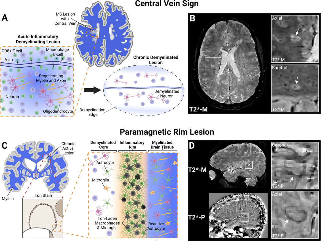
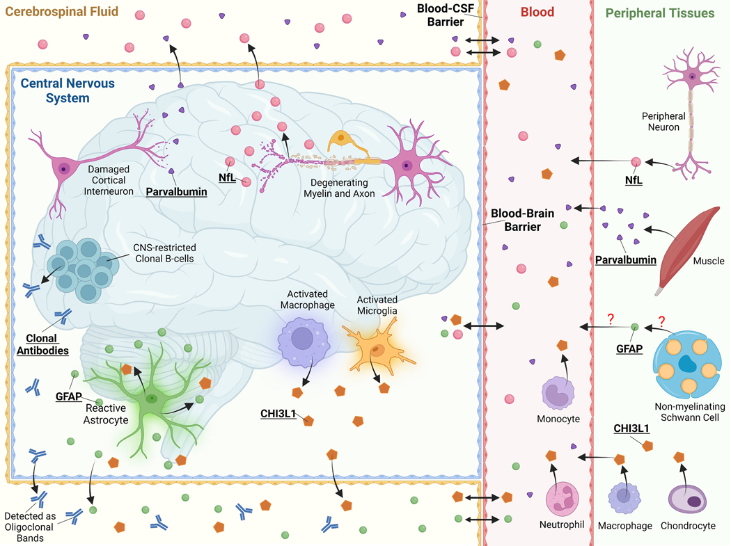
The advent of highly effective disease modifying therapy has transformed the landscape of multiple sclerosis (MS) care over the last two decades. However, there remains a critical, unmet need for sensitive and specific biomarkers to aid in diagnosis, prognosis, treatment monitoring, and the development of new interventions, particularly for people with progressive disease. This review evaluates the current data for several emerging imaging and liquid biomarkers in people with MS. MRI findings such as the central vein sign and paramagnetic rim lesions may improve MS diagnostic accuracy and evaluation of therapy efficacy in progressive disease. Serum and cerebrospinal fluid levels of several neuroglial proteins, such as neurofilament light chain and glial fibrillary acidic protein, show potential to be sensitive biomarkers of pathologic processes such as neuro-axonal injury or glial-inflammation. Additional promising biomarkers, including optical coherence tomography, cytokines and chemokines, microRNAs, and extracellular vesicles/exosomes, are also reviewed, among others. Beyond their potential integration into MS clinical care and interventional trials, several of these biomarkers may be informative of MS pathogenesis and help elucidate novel targets for treatment strategies.
Keywords: Central vein sign; Glial fibrillary acidic protein; Neurofilament light chain; Optical coherence tomography; Paramagnetic rim lesion.
© 2023 Wiley-VCH GmbH.
Conflict of interest statement
Conflicts of Interest Disclosure:
AJG and SPG have no conflicts of interest.
Figures
Similar articles
-
The Black Book of Psychotropic Dosing and Monitoring.Psychopharmacol Bull. 2024 Jul 8;54(3):8-59. Psychopharmacol Bull. 2024. PMID: 38993656 Free PMC article. Review.
-
Neurofilament light chain and glial fibrillary acidic protein as diagnostic and prognostic biomarkers in epileptic seizures and epilepsy: A systematic review.Epilepsy Behav. 2025 Apr;165:110321. doi: 10.1016/j.yebeh.2025.110321. Epub 2025 Feb 20. Epilepsy Behav. 2025. PMID: 39983592
-
Current and Future Biomarkers in Multiple Sclerosis.Int J Mol Sci. 2022 May 24;23(11):5877. doi: 10.3390/ijms23115877. Int J Mol Sci. 2022. PMID: 35682558 Free PMC article. Review.
-
Uncommon Non-MS Demyelinating Disorders of the Central Nervous System.Curr Neurol Neurosci Rep. 2025 Jul 1;25(1):45. doi: 10.1007/s11910-025-01432-8. Curr Neurol Neurosci Rep. 2025. PMID: 40591029 Review.
-
Glial cell injury and atrophied lesion volume as measures of chronic multiple sclerosis inflammation.J Neurol Sci. 2024 Jun 15;461:123055. doi: 10.1016/j.jns.2024.123055. Epub 2024 May 14. J Neurol Sci. 2024. PMID: 38761669
Cited by
-
Multiple Sclerosis: From the Application of Oligoclonal Bands to Novel Potential Biomarkers.Int J Mol Sci. 2024 May 15;25(10):5412. doi: 10.3390/ijms25105412. Int J Mol Sci. 2024. PMID: 38791450 Free PMC article. Review.
-
Proteomic Biomarker Panel for Gauging Multiple Sclerosis Disease Activity: A Case Series From Real-World Use.Int J MS Care. 2025 Mar 24;27(Q1):90-94. doi: 10.7224/1537-2073.2024-094. eCollection 2025 Jan. Int J MS Care. 2025. PMID: 40135190 Free PMC article.
-
Multi-organ imaging-derived polygenic indexes for brain and body health.medRxiv [Preprint]. 2024 Jun 3:2023.04.18.23288769. doi: 10.1101/2023.04.18.23288769. medRxiv. 2024. PMID: 38883759 Free PMC article. Preprint.
-
Biomarkers for neurodegenerative diseases in regulatory decision-making by the European Medicines Agency.Alzheimers Dement (N Y). 2025 Mar 27;11(1):e70072. doi: 10.1002/trc2.70072. eCollection 2025 Jan-Mar. Alzheimers Dement (N Y). 2025. PMID: 40151396 Free PMC article.
-
Glial Fibrillary Acidic Protein as a Marker of Disease in Relapsing Multiple Sclerosis: Post Hoc Analysis of Phase 3 Ozanimod Trials.Eur J Neurol. 2025 Jun;32(6):e70222. doi: 10.1111/ene.70222. Eur J Neurol. 2025. PMID: 40462564 Free PMC article. Clinical Trial.
References
-
- Yamout BI, Khoury SJ, Ayyoubi N, et al. Alternative diagnoses in patients referred to specialized centers for suspected MS. Mult Scler Relat Disord 2017;18:85–89. - PubMed
-
- Kaisey M, Solomon AJ, Luu M, Giesser BS, Sicotte NL. Incidence of multiple sclerosis misdiagnosis in referrals to two academic centers. Mult Scler Relat Disord 2019;30:51–56. - PubMed
-
- Ascherio A, Munger KL. Environmental risk factors for multiple sclerosis. Part I: the role of infection. Ann Neurol 2007;61(4):288–99. - PubMed
Publication types
MeSH terms
Substances
Grants and funding
LinkOut - more resources
Full Text Sources
Medical
