

Review
doi: 10.1259/bjr.20221096.
Epub 2023 May 25.
Radiological features of nephrocalcinosis, a common but forgotten entity
Affiliations
- PMID: 37194990
- PMCID: PMC10392641
- DOI: 10.1259/bjr.20221096
Item in Clipboard
Review
Radiological features of nephrocalcinosis, a common but forgotten entity
Br J Radiol.
2023 Aug.
Abstract
Nephrocalcinosis refers to calcium deposition in the form of calcium oxalate or calcium phosphate in the renal parenchyma and tubules. After diagnosis, the cause of nephrocalcinosis must be established to carry out a comprehensive approach to this entity. Although this is a common finding, it can be underdiagnosed due to the lack of knowledge of the different presentation patterns that exist. Many causes have been described related to this disease.A pictorial review about the most common features of cortical and medullary nephrocalcinosis both in ultrasound and CT is presented in the present work as well as a review of its main causes and graphics to easily recognize each pattern.
Figures

Axial Non-contrast CT section of the kidneys: linear or “band” cortical nephrocalcinosis pattern. Bilateral cortical linear hiperdensities.

Illustration of cortical nephrocalcinosis patterns: a. Linear or Banded, b: Punctate, c: Banded: “tram sign”.

(a) Axial non-contrast CT section of the right kidney and (b) axial non-contrast CT section of the left kidney: medullary nephrocalcinosis, with peripheral hyperechogenic band with central involvement pattern, the arrows indicate a peripheral calcification of the renal pyramid.

Renal ultrasound: (a) sagittal slice and (b) coronal slice: Medullary nephrocalcinosis, showcasing a global increase in the echogenicity of the pyramid, the marked acoustic shadow can be distinguished.

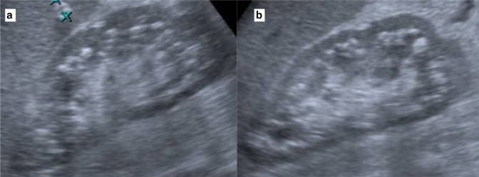
Renal ultrasound. (a) coronal slice and (b) sagittal slice: Medullary nephrocalcinosis. The arrows show global increase in echogenicity of the pyramid, without generating posterior acoustic shadow.

Axial non-contrast CT section of the right kidney, Medullary nephrocalcinosis, Anderson-Carr-Randal pattern showing a linear echogenic focus at the tip of the renal pyramid along the margins of the fornix.

Illustration patterns of medullary nephrocalcinosis presentation: (a) linear without compromising the papilla, (b) linear with more hyperechogenic in the middle of the renal pyramid, (c) global involvement of the pyramid, (d) Anderson-Carr-Randall pattern, calcification in the papilla and towards the fornixes.
(a, b) Renal sagittal ultrasound slices showing mixed pattern of medullary nephrocalcinosis with involvement of the renal pyramids and fornix.

Renal sagittal ultrasound slice showing medullary nephrocalcinosis, Anderson-Carr-Randall pattern: calcification towards the renal papilla is seen.
References
-
- Vaidya SR, Yarrarapu SNS, Aeddula NR. Nephrocalcinosis. In: Statpearls [ Internet ] Treasure Island (Fl): Statpearls Publishing 2021.
Publication types
MeSH terms
Substances
LinkOut - more resources
Full Text Sources
