. 2023 Jul;44(10):4101-4119.
doi: 10.1002/hbm.26333. Epub 2023 May 17.

Brain-wide associations between white matter and age highlight the role of fornix microstructure in brain ageing

Affiliations

Brain-wide associations between white matter and age highlight the role of fornix microstructure in brain ageing

Max Korbmacher et al. Hum Brain Mapp. 2023 Jul.

Abstract

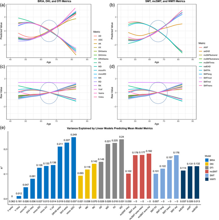
Unveiling the details of white matter (WM) maturation throughout ageing is a fundamental question for understanding the ageing brain. In an extensive comparison of brain age predictions and age-associations of WM features from different diffusion approaches, we analyzed UK Biobank diffusion magnetic resonance imaging (dMRI) data across midlife and older age (N = 35,749, 44.6-82.8 years of age). Conventional and advanced dMRI approaches were consistent in predicting brain age. WM-age associations indicate a steady microstructure degeneration with increasing age from midlife to older ages. Brain age was estimated best when combining diffusion approaches, showing different aspects of WM contributing to brain age. Fornix was found as the central region for brain age predictions across diffusion approaches in complement to forceps minor as another important region. These regions exhibited a general pattern of positive associations with age for intra axonal water fractions, axial, radial diffusivities, and negative relationships with age for mean diffusivities, fractional anisotropy, kurtosis. We encourage the application of multiple dMRI approaches for detailed insights into WM, and the further investigation of fornix and forceps as potential biomarkers of brain age and ageing.

Keywords: ageing; brain age; diffusion; forceps; fornix; magnetic resonance imaging; white matter.


Conflict of interest statement

OOA has received a speaker's honorarium from Lundbeck and is a cosultant to Coretechs.ai.

Figures

FIGURE 1
FIGURE 1
Density plots for the sample's age by sex and scanner site. The y‐axis indicates the probability of age scaled to 1.
FIGURE 2
FIGURE 2
Model performance for different train‐test splits. Model metrics R 2, root mean squared error (RMSE), mean absolute error (MAE) and their standard deviations, as well as the Pearson's correlations between predicted and chronological age and its 95% confidence interval are displayed for different training data percentages of the total data (x‐axis). For visualization purposes, RMSE and MAE were divided by 10. For exact values see Table S1.
FIGURE 3
FIGURE 3
Corrected and uncorrected brain age by age for each of the utilized brain age models.
FIGURE 4
FIGURE 4
Overview of the analysis steps.
FIGURE 5
FIGURE 5
Differences between Pearson's correlations of chronological and uncorrected predicted ages across diffusion approaches with 95% confidence interval. Differences between Pearson's correlation coefficients of chronological and uncorrected predicted age by diffusion approach. See Figure S8 for correlational differences between approaches for corrected brain age predictions.
FIGURE 6
FIGURE 6
Correlations of uncorrected BAG and age across used diffusion approaches. Age‐BAG correlations were significant at pHolm < .001. For the corrected BAG correlations across models see Figure S1.
FIGURE 7
FIGURE 7
Correlation matrix for fornix diffusion metrics and chronological age. All correlations were significant at Holm‐corrected pHolm < .05.
FIGURE 8
FIGURE 8
Correlations between diffusion metrics and age. Each point indicates one correlation between a diffusion metric and chronological age. Names of diffusion metrics are displayed when correlations between the metric and age reached a Pearson correlation of |r| > 0.5. Holm correction (Holm, 1979) was used for Holm‐correction, and all displayed values were significant at p < .001. For the distribution of the correlations see Figure S12.
FIGURE 9
FIGURE 9
Whole‐brain and fornix diffusion metrics across age. The presented plots represent diffusion metrics for each of the six diffusion models from the full sample N = 35,749 for (a) whole‐brain diffusion metrics, (b) fornix diffusion metrics. Brighter colors indicate higher density and red lines are fitted lines to the relationship between age and diffusion metric. Plots for forceps can be found in Figure S14.
FIGURE 10
FIGURE 10
Raw and predicted whole‐brain WM diffusion metrics by chronological age. Figure 10a–d shows age curves for each standardized (z‐score) diffusion metric's mean skeleton value (y‐axis) plotted as a function of age (x‐axis). Shaded areas represent 95% CI. Curves fitted to raw values (Figure 10c,d) serve as a comparison to the lm‐derived predicted values from Equation (4) (Figure a,b). Figure 10e indicates the model fit for the linear models from Figure 10a,b, showing R 2adj values on top and standard error (SE) on the bottom of the bars which each represent a Fornix skeleton value for one of the seven models. Lines crossing at age 65 are marked with ovals. Model summaries of all 28 mean models can be found in Table S5. The same visualization of fornix diffusion values can be found in Figure S9, and for the forceps minor in Figure S15.

References

    1. Alzheimer's Association. (2020). Alzheimer's disease facts and figures. Alzheimer's & Dementia, 16, 391–460. - PubMed
    1. Andersson, J. L. R. , & Sotiropoulos, S. N. (2016). An integrated approach to correction for off‐resonance effects and subject movement in diffusion MR imaging. NeuroImage, 125, 1063–1078. - PMC - PubMed
    1. Bach, M. , Laun, F. B. , Leemans, A. , Tax, C. M. W. , Biessels, G. J. , Stieltjes, B. , & Maier‐Hein, K. H. (2014). Methodological considerations on tract‐based spatial statistics (TBSS). NeuroImage, 100, 358–369. - PubMed
    1. Baecker, L. , Garcia‐Dias, R. , Vieira, S. , Scarpazza, C. , & Mechelli, A. (2021). Machine learning for brain age prediction: Introduction to methods and clinical applications. eBioMedicine, 72, 103600. - PMC - PubMed
    1. Barrick, T. R. , Charlton, R. A. , Clark, C. A. , & Markus, H. S. (2010). White matter structural decline in normal ageing: A prospective longitudinal study using tract‐based spatial statistics. NeuroImage, 51, 565–577. - PubMed

Publication types