. 2023 Sep;48(9):2857-2869.
doi: 10.1007/s11064-023-03952-6. Epub 2023 May 17.

Functional Analysis of a Novel Immortalized Murine Microglia Cell Line in 3D Spheroid Model

Affiliations

Functional Analysis of a Novel Immortalized Murine Microglia Cell Line in 3D Spheroid Model

Gabrielle Angst et al. Neurochem Res. 2023 Sep.

Abstract

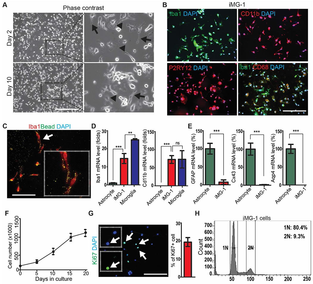
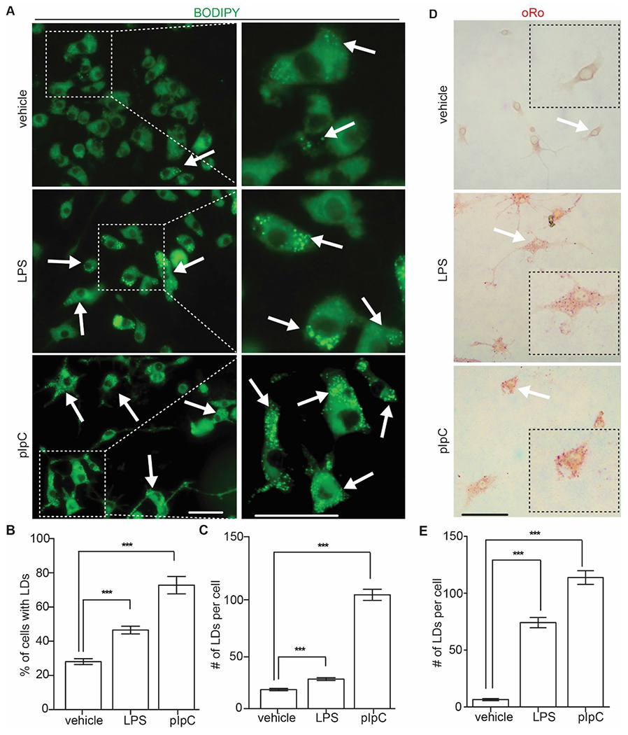
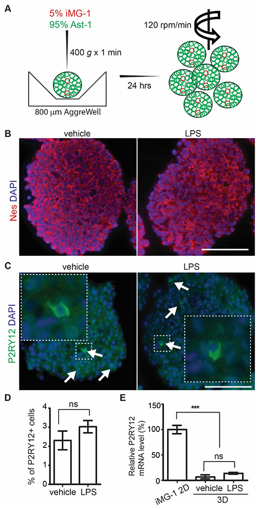
Microglia are the residential immune cells of central nervous system and they are crucial for brain development and homeostasis, as well as the progression of inflammatory brain diseases. To study microglia's physiological and pathological functions, one of the most widely used models is primary microglia culture from neonatal rodents. However, primary microglia culture is time consuming and needs a great number of animals. In our microglia culture, we found a strain of spontaneously immortalized microglia that continued to divide without any known genetic intervention. We confirmed the immortalization of these cells for uninterrupted thirty passages and we named them as immortalized microglia like-1 cells (iMG-1). The iMG-1 cells kept their microglia morphology, and they expressed macrophage/microglia-specific proteins of CD11b, CD68, P2RY12, and IBA1 in vitro. iMG-1 cells were responsive to inflammatory stimulations with lipopolysaccharide (LPS) and Polyinosinic:polycytidylic acid (pIpC), triggering increased mRNA/protein levels of IL1-β, IL-6, TNF-α, and interferons. LPS and pIpC treated iMG-1 cells also significantly increased their accumulation of lipid droplets (LDs). We also generated a 3D spheroid model using immortalized neural progenitor cells and iMG-1 cells with defined percentages to study neuroinflammation. The iMG-1 cells distributed evenly in spheroids, and they regulated the basal mRNA levels of cytokines of neural progenitors in 3D spheroid. iMG-1 cells were responsive to LPS by increased expression of IL-6 and IL1-β in spheroids. Together, this study indicated the reliability of iMG-1 which could be readily available to study the physiological and pathological functions of microglia.

Keywords: Coculture; Immortalization; LPS; Microglia; Neuroinflammation; Spheroids; pIpC.


Conflict of interest statement

Conflict of interest The authors declare no conflicts of interest.

Figures

Fig. 1
Fig. 1
Establishment of a microglia-like iMG-1 cell line. A Representative phase contrast images of iMG-1 cells after 2 and 10 days in culture. The boxed areas were shown in detail on the right. B Immunofluorescence of IBA1, CD11B, P2RY12, CD68 and DAPI in iMG-1 cells. C Immunofluorescence of IBA1 and phagocytosed fluorescent beads in iMG-1 cells. The arrow indicated cells were enlarged to show as inset. D and E Mean ± S.E. of relative mRNA expression levels of Iba1 and Cd11b D, GFAP, Cx34 and Aqp4 E in primary astrocyte, primary microglia, and iMG-1 cells were shown. F Mean ± S.E. of cell number of iMG-1 cells for 0, 5, 10, 15, and 20 days in culture were shown. Experiments were repeated 5–6 times for each time point. G Immunofluorescence of Ki67 and DAPI in iMG-1 cells 3 days after reseeding. Mean ± S.E. of the percentage of Ki67+ iMG-1 cells were shown. H Cell cycle analysis of iMG-1 cells by FACS. Two-way anova and tukey post-hoc tests were used for statistic tests. Ns no significance, **p < 0.01, ***p < 0.001. Bar = 100 μm
Fig. 2
Fig. 2
Proinflammation of iMG-1 cells in response to LPS and pIpC stimulations. A Representative phase contrast images of iMG-1 cells after treatment of vehicle, LPS and pIpC for 24 h. B and C Mean ± S.E. of relative mRNA expression levels of IL-6, TNF-α, IL-1β, IFN-α, IFN-β, and IFN-γ B, Arg-1 and YM-1 C in iMG-1 cells after vehicle, LPS, or pIpC treatment were shown. D Expression levels of TNF-α and IL-6 by FACS analysis of iMG-1 cells after treatments of vehicle, LPS, or pIpC for 24 h. Experiments were repeated 4–6 times. Ns no significance, *p < 0.05, **p < 0.01, ***p < 0.001. Bar = 50 μm
Fig. 3
Fig. 3
LDs accumulation in LPS and pIpC treated iMG-1 cells. A Fluorescence of LDs in iMG-1 cells after vehicle, LPS, or pIpC treatment. Arrows indicated BODIPY+ iMG-1 cells. The boxed areas were shown in detail on the right. B Mean ± S.E. of percentage of BODIPY+ iMG-1 cells after vehicle, LPS, or pIpC treatment were shown. C Mean ± S.E. of LDs number of each BODIPY+ iMG-1 cells after vehicle, LPS, or pIpC treatment were shown. D oRo staining of LDs in iMG-1 cells after vehicle, LPS, or pIpC treatment. E Mean ± S.E. of LDs number of each oRo+ iMG-1 cells after vehicle, LPS, or pIpC treatment were shown. For BODIPY staining, 709 vehicle treated cells, 785 LPS treated cells, and 417 pIpC treated cells were counted. For oRo staining, 97 vehicle treated cells, 107 LPS treated cells, and 121 pIpC treated iMG-1 cells were counted. ***p < 0.001. Bar = 10 μm
Fig. 4
Fig. 4
Incorporation of iMG-1 cells into 3D neural spheroid culture. A Schematic graphs of 3D spheroids generation using iMG-1 cells and Ast-1 cells. B, C Immunofluorescence of nestin and DAPI B, P2RY12 and DAPI C of iMG-1 cells in 3D spheroid coculture after vehicle or LPS treatment. Arrows in C indicated P2RY12+ iMG-1 cells in spheroid. The boxed areas were shown in detail as insets. D Mean ± S.E. of the percentage of P2RY12+ cell of cocultured spheroid cell after vehicle and LPS treatment were shown. E Mean ± S.E. of relative mRNA expression level of P2ry12 in cocultured spheroid after vehicle and LPS treatment were shown. Experiments were repeated 5–6 times. Ns no significance, ***p < 0.001. Bar = 100 μm
Fig. 5
Fig. 5
Altered cytokine expression by inoculating iMG-1 cells in 3D spheroid culture. Mean ± S.E. of relative mRNA expression levels of IL-1β, IL-6, TNF-α, IFN-α, IFN-β, and IFN-γ in cultured spheroids with or without iMG-1 cell inoculation after vehicle and LPS treatment were shown. Experiments were repeated 5–6 times. Ns no significance, *p < 0.05, **p < 0.01, ***p < 0.001

References

    1. Ginhoux F et al. (2010) Fate mapping analysis reveals that adult microglia derive from primitive macrophages. Science 330(6005):841–845 - PMC - PubMed
    1. Li Q, Barres BA (2018) Microglia and macrophages in brain homeostasis and disease. Nat Rev Immunol 18(4):225–242 - PubMed
    1. Wolf SA, Boddeke HW, Kettenmann H (2017) Microglia in physiology and disease. Annu Rev Physiol 79:619–643 - PubMed
    1. Saijo K, Glass CK (2011) Microglial cell origin and phenotypes in health and disease. Nat Rev Immunol 11(11):775–787 - PubMed
    1. Schafer DP, Stevens B (2015) Microglia function in central nervous system development and plasticity. Cold Spring Harb Perspect Biol 7(10):a020545. - PMC - PubMed