

EEG Microstates as a Signature of Hemispheric Lateralization in Stroke
- PMID: 37195492
- PMCID: PMC10191079
- DOI: 10.1007/s10548-023-00967-8
EEG Microstates as a Signature of Hemispheric Lateralization in Stroke
Abstract
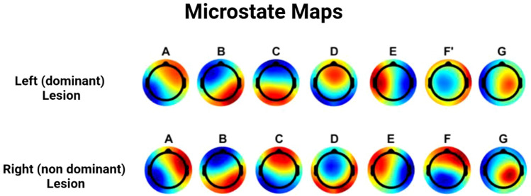
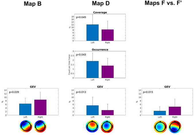
Stroke recovery trajectories vary substantially. The need for tracking and prognostic biomarkers in stroke is utmost for prognostic and rehabilitative goals: electroencephalography (EEG) advanced signal analysis may provide useful tools toward this aim. EEG microstates quantify changes in configuration of neuronal generators of short-lasting periods of coordinated synchronized communication within large-scale brain networks: this feature is expected to be impaired in stroke. To characterize the spatio-temporal signatures of EEG microstates in stroke survivors in the acute/subacute phase, EEG microstate analysis was performed in 51 first-ever ischemic stroke survivors [(28-82) years, 24 with right hemisphere (RH) lesion] who underwent a resting-state EEG recording in the acute and subacute phase (from 48 h up to 42 days after the event). Microstates were characterized based on 4 parameters: global explained variance (GEV), mean duration, occurrences per second, and percentage of coverage. Wilcoxon Rank Sum tests were performed to compare features of each microstate across the two groups [i.e., left hemisphere (LH) and right hemisphere (RH) stroke survivors]. The canonical microstate map D, characterized by a mostly frontal topography, displayed greater GEV, occurrence per second, and percentage of coverage in LH than in RH stroke survivors (p < 0.05). The EEG microstate map B, with a left-frontal to right-posterior topography, and F, with an occipital-to-frontal topography, exhibited a greater GEV in RH than in LH stroke survivors (p = 0.015). EEG microstates identified specific topographic maps which characterize stroke survivors' lesioned hemisphere in the acute and early subacute phase. Microstate features offer an additional tool to identify different neural reorganization.
Keywords: EEG; Hemispheric lateralization; Microstates; Stroke.
© 2023. The Author(s).
Conflict of interest statement
The authors declare they have no conflict of interest.
Figures
References
-
- Bentes C, Peralta AR, Viana P, Martins H, Morgado C, Casimiro C, Franco AC, Fonseca AC, Geraldes R, Canhão P, Pinho E, Melo T, Paiva T, Ferro JM. Quantitative EEG and functional outcome following acute ischemic stroke. Clin Neurophysiol. 2018;129(8):1680–1687. doi: 10.1016/j.clinph.2018.05.021. - DOI - PubMed
MeSH terms
Grants and funding
- REACT EU-PON Ricerca e Innovazione 2014-2020, DM 1062/2021/European Commission
- C96C18000370001/Italian Ministry of Education (MIUR)
- H2020 Research and Innovation Staff Exchange grant: PRO GAIT grant agreement No. 778043/EU
- PGR-01045 (SoftAct project)/Italian Ministry for foreign Affairs and International Cooperation
- UNIPD research funding/Università degli Studi di Padova
LinkOut - more resources
Full Text Sources
Medical
