. 2023 May 1;7(5):333-352.
doi: 10.4049/immunohorizons.2300013.

B Cell Chronic Lymphocytic Leukemia Development in Mice with Chronic Lung Exposure to Coccidioides Fungal Arthroconidia

Affiliations

B Cell Chronic Lymphocytic Leukemia Development in Mice with Chronic Lung Exposure to Coccidioides Fungal Arthroconidia

Vanessa Coyne et al. Immunohorizons. .

Abstract

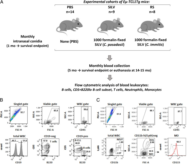
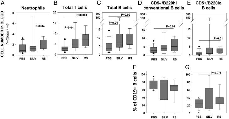
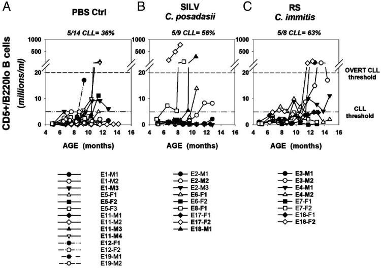
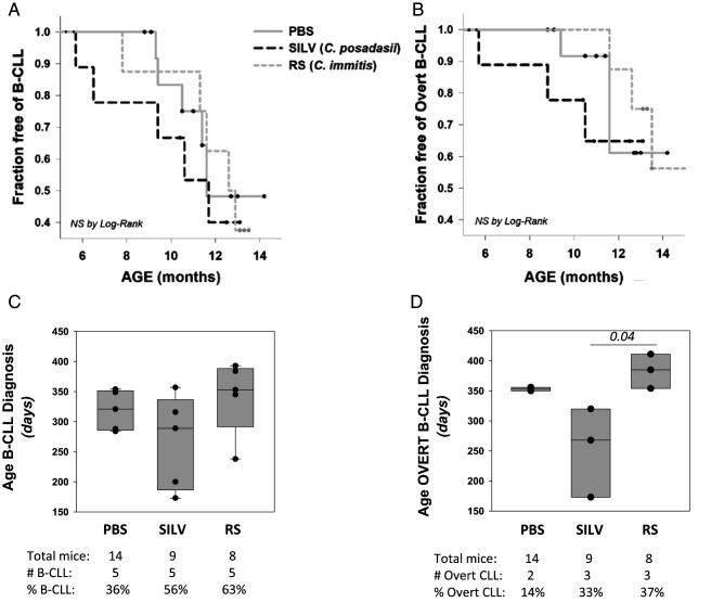
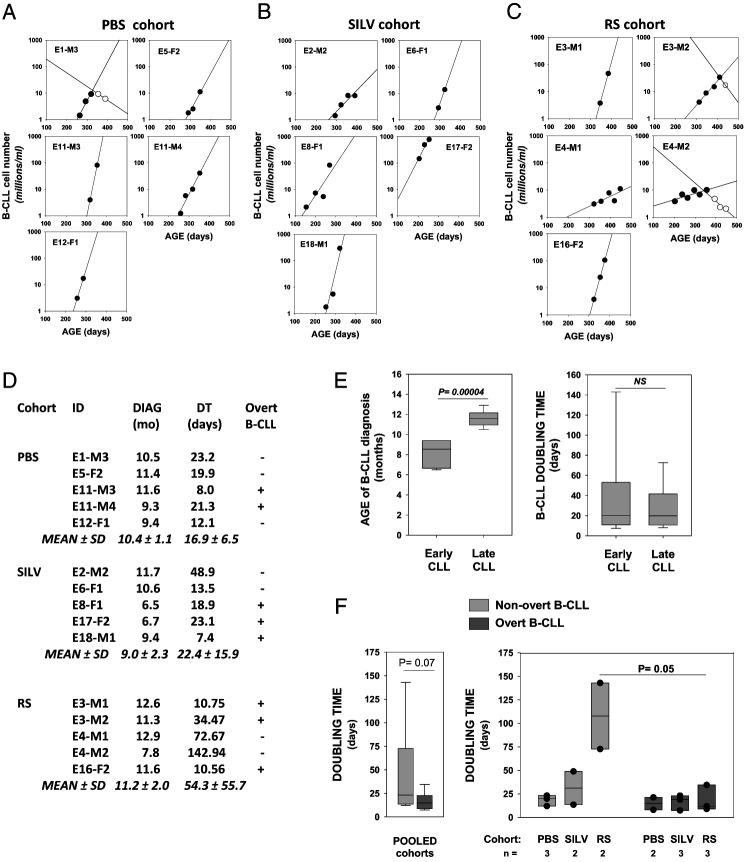
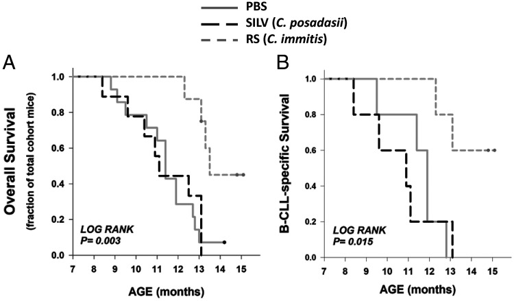
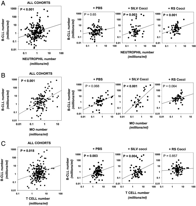
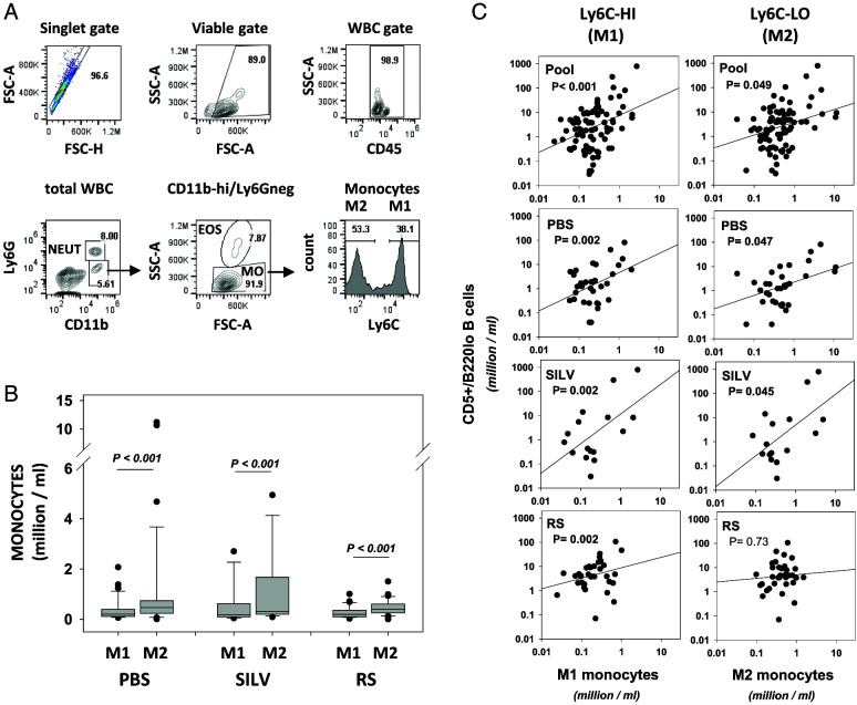
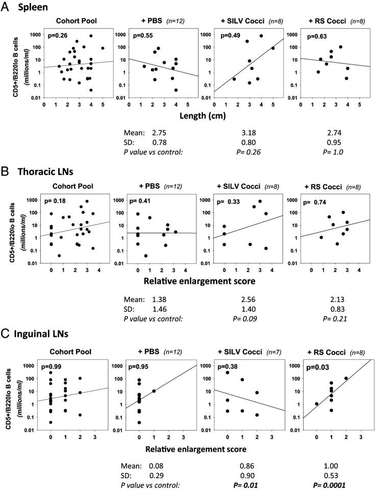
Links between repeated microbial infections and B cell chronic lymphocytic leukemia (B-CLL) have been proposed but not tested directly. This study examines how prolonged exposure to a human fungal pathogen impacts B-CLL development in Eµ-hTCL1-transgenic mice. Monthly lung exposure to inactivated Coccidioides arthroconidia, agents of Valley fever, altered leukemia development in a species-specific manner, with Coccidioides posadasii hastening B-CLL diagnosis/progression in a fraction of mice and Coccidioides immitis delaying aggressive B-CLL development, despite fostering more rapid monoclonal B cell lymphocytosis. Overall survival did not differ significantly between control and C. posadasii-treated cohorts but was significantly extended in C. immitis-exposed mice. In vivo doubling time analyses of pooled B-CLL showed no difference in growth rates of early and late leukemias. However, within C. immitis-treated mice, B-CLL manifests longer doubling times, as compared with B-CLL in control or C. posadasii-treated mice, and/or evidence of clonal contraction over time. Through linear regression, positive relationships were noted between circulating levels of CD5+/B220low B cells and hematopoietic cells previously linked to B-CLL growth, albeit in a cohort-specific manner. Neutrophils were positively linked to accelerated growth in mice exposed to either Coccidioides species, but not in control mice. Conversely, only C. posadasii-exposed and control cohorts displayed positive links between CD5+/B220low B cell frequency and abundance of M2 anti-inflammatory monocytes and T cells. The current study provides evidence that chronic lung exposure to fungal arthroconidia affects B-CLL development in a manner dependent on fungal genotype. Correlative studies suggest that fungal species differences in the modulation of nonleukemic hematopoietic cells are involved.


Conflict of interest statement

The authors have no financial conflicts of interest.

Figures

FIGURE 1.
FIGURE 1.
Experimental design and gating strategy for diverse blood leukocytes. (A) With three cohorts of B-CLL–prone TCL1-Tg mice, we examined whether B-CLL development is affected by repeated inhalation of Coccidioides arthroconidia. The control cohort was exposed to vehicle alone (PBS; n = 14); test cohorts received 1000 fixed C. posadasii conidia (SILV; n = 9) or 1000 fixed C. immitis conidia (RS; n = 8) monthly, beginning at ∼1 mo of age and continuing throughout each recipient’s lifespan. Blood samples were acquired monthly, beginning at 5–6 mo. Total WBCs in individual samples were counted and normal and leukemic cells were distinguished by immunofluorescence staining and flow cytometry. (B) Gating strategy for B and T lymphocytes. Viable singlets were gated for the pan-leukocyte marker, CD45, and assessed for CD19 expression. T cells within the CD19 population were identified by their CD5high status. Normal and leukemic B cells within the CD19+ gate were distinguished as follows: B-CLL cells, CD5+/B220low; normal B cells, CD5/B220high. (C) Neutrophils and monocytes. Viable singlets, gated for CD45, were evaluated for CD11b and Ly6G expression. Neutrophils are Ly6G+/CD11bhigh; monocyte-enriched WBCs are Ly6G/CD11bhigh. Contaminating eosinophils were removed from the latter by gating out cells with high side scatter (SSC). The resulting monocyte population (Ly6G/CD11bhigh/SSClow) expressed CD115, a marker of blood monocytes. Note that in most mice ≤7 mo of age, monocytes were not enumerated due to suboptimal monocyte staining/gating, and neutrophils were gated either as CD45+CD19CD5/SSChigh or as CD45+/Ly6G+. Later comparisons indicated that the latter two approaches at neutrophil enumeration yielded results highly concordant with neutrophils gated as CD45+/CD11bhigh/Ly6G+ (C).
FIGURE 2.
FIGURE 2.
Coccidioides lung exposure alters blood levels of several leukocyte populations at 6–7 mo of age. Shown is the cell frequency (millions/ml) of blood leukocyte populations in mice of each cohort: PBS (n = 14 mice), SILV (C. posadasii treated; n = 9), or RS (C. immitis treated; n = 8). (AE) Boxplots show frequencies of (A) neutrophils, (B) total T cells, (C) total B cells, (D) nonleukemic, normal B cells (CD5/B220high), and (E) cells with a preleukemic/leukemic phenotype (CD5+/B220low). Note that due to suboptimal monocyte staining in most blood samples taken at this age, insufficient data were available for statistical comparisons of monocyte numbers between cohorts. Nonetheless for two mice of each cohort, satisfactorily stained/gated for monocytes (as in Fig. 1C), the following frequencies were noted (mean ± SD in millions/ml): PBS, 0.24 ± 0.27; SILV, 1.38 ± 1.19; and RS, 0.43 ± 0.04. (F) Percent of total CD19+ cells with normal B cell phenotype and (G) percent of total CD19+ cells with leukemic phenotype. Differences were determined as statistically significant using a t test, for data with a normal distribution, or a Mann–Whitney rank-sum test for nonparametric data.
FIGURE 3.
FIGURE 3.
Time course for B-CLL emergence within PBS- and Coccidioides-exposed TCL1-Tg mice. (AC) Shown are blood frequencies of B cells with the leukemic phenotype (CD5+/B220low) over the lifespan of individual mice chronically exposed to (A) PBS, (B) SILV, or (C) RS. The lower reference line within each graph represents the threshold frequency for B-CLL diagnosis (5 million/ml; see Materials and Methods for added criteria). The upper reference line represents the threshold for overt B-CLL diagnosis (20 million CD5+/B220low B cells/ml). The legend below each graph supplies mouse identification codes; those in bold achieved a B-CLL diagnosis.
FIGURE 4.
FIGURE 4.
Statistical analysis of B-CLL incidence with age. (A) Kaplan–Meier incidence curves, representing the percentage of each cohort that is B-CLL–free over time. Small filled circles stand for censored events, reflecting mouse death without a B-CLL diagnosis. (B) Kaplan–Meier incidence curve, representing the percentage of each cohort that is free of overt B-CLL over time. PBS cohort, solid gray; SILV, dashed black; RS, short dash gray. Differences observed in (A) or (B) plots did not reach statistical significance by log-rank analysis. (C) Boxplot analysis of pooled data representing age at B-CLL diagnosis within each cohort. Boxes show median levels, with upper and lower quartiles and whiskers representing variability outside the quartiles. Overlaid black circles represent values of individual mice. Statistical analysis (Kruskal–Wallis one-way ANOVA on ranks) showed no significant difference between these groups (p = 0.491). (D) Boxplot of pooled data representing age at overt B-CLL diagnosis within each cohort. Statistical analysis (as above) revealed a statistically significant difference between these groups (p = 0.025). B-CLL cases in the PBS cohort were insufficient to make pairwise comparisons with other cohorts through the unpaired, two-sided t test. Nonetheless, ages for overt B-CLL diagnosis in the SILV cohort were statistically different from those in the RS cohort (p = 0.03) by the latter analysis.
FIGURE 5.
FIGURE 5.
Doubling time determinations for B-CLL populations in control and Coccidioides-exposed mice. (AC) Linear regression plots showing frequency of CD5+/B220low B cells (log) versus age of mice diagnosed with B-CLL. (A) PBS. (B) SILV. (C) (RS). Lines with filled symbols show the progressive increase in B-CLL numbers over time. The frequencies of CD5+/B220low cells that qualify as preleukemic monoclonal B cell lymphocytosis (MBL = 0.5–5 million/ml) were included in these plots when the latter supplied added confidence in the linear growth curve. Contraction of the leukemic population was seen in certain mice (E1-M3 in PBS cohort; E3-M2 and E4-M2 in RS cohort). Leukemic cell counts during contraction are shown by open symbols; these were not used to compute initial B-CLL doubling time (DT). (D) Age of diagnosis (months), DT values (days), and overt status of each B-CLL within the PBS, SILV, and RS cohorts. Calculation of DT was achieved with an online DT calculator based on exponential regression (http://www.doubling-time.com/compute). (E) Pooled B-CLLs were categorized as early CLL (n = 6; range, 6.5–9.4 mo) or late CLL (n = 9; range, 10.5–12.9 mo) based on age at diagnosis. By boxplot analysis, the two groups were compared for statistically significant differences in (left) age of B-CLL diagnosis (p = 0.00004 by a two-sided, unpaired t test) and (right) B-CLL DT (not significant). (F) Left, Boxplot analysis for DT in total overt versus total nonovert B-CLL populations (p = 0.070 by nonparametric Mann–Whitney rank-sum test). Boxplots for B-CLL DTs in nonovert (light gray bars) versus overt B-CLL (dark gray bars) of each experimental cohort (overlaid filled symbols represent individual mouse values). Median DT for overt B-CLL was consistently low in all cohorts (no statistically significant difference by Kruskal–Wallis one-way ANOVA on ranks). Greater intercohort diversity in DT was seen in nonovert B-CLL, without reaching statistical significance. Nonetheless, within the RS cohort, DT values for nonovert and overt B-CLL were significantly different (p = 0.025 by a one-sided and p = 0.05 by a two-sided, unpaired t test).
FIGURE 6.
FIGURE 6.
Overall and disease-specific survival times in control and Coccidioides-exposed TCL1-Tg mice. (A) Kaplan–Meier incidence curves for overall survival (PBS cohort, solid gray; SILV, dashed black; RS, short dash gray). Log rank analysis showed a statistically significant difference within the set of three cohorts (p = 0.003). Statistical significance for all pairwise comparisons is as follows (Holm–Sidak test): PBS versus RS (p = 0.004), SILV versus RS (p = 0.004), and PBS versus SILV (p = 0.717). (B) Kaplan-Meier incidence curves for disease-specific survival (all mice in this analysis developed B-CLL during the 14-mo course of this study). Log rank analysis showed a statistically significant difference within the set of three cohorts (p = 0.015; Holm–Sidak test for pairwise comparisons): PBS versus RS (p = 0.02), SILV versus RS (p = 0.03), and PBS versus SILV (p = 0.18).
FIGURE 7.
FIGURE 7.
CD5+/B220low (preleukemic and leukemic) cell levels in blood are linked to the frequency of other blood leukocyte populations in a cohort-specific manner. (AC) Frequency of CD5+/B220low B cells detected in individual mice at any one blood sampling was plotted against the respective frequency of either (A) neutrophils, (B) monocytes, or (C) T cells. Dot plots and accompanying linear regression lines are shown for pooled mice and mice of each experimental cohort. Values represent data from sequential monthly blood analyses, beginning at 5–6 mo to survival endpoint. In the case of monocytes, fewer mice of each cohort were measured at 5-6 mo. The p values for each linear regression analysis are shown, with those reaching statistical significance (p < 0.05) in bold.
FIGURE 8.
FIGURE 8.
Circulating levels of CD5+/B220low B cells are significantly correlated with the frequency of inflammatory (M1) and anti-inflammatory (M2) monocyte subsets in a cohort-specific manner. (A) Gating strategy for segregating classical, inflammatory (M1) monocytes and nonclassical, anti-inflammatory (patrolling) (M2) monocytes. M1 cells represent monocytes (CD45+/Ly6G/CD11bhigh/SSClow) that are gated as Ly6Chigh. M2 cells represent monocytes gated as Ly6C. Note that monocytes bearing intermediate levels of Ly6C, thought to represent transitional monocytes (64, 65), are not considered. (B) Boxplots showing M1 and M2 cell frequencies in pooled monthly bleeds from control TCL1-Tg mice and similar blood samples from SILV-treated and RS-treated cohorts (PBS cohort, n = 13 mice represented with 34 samplings during 6–13 mo; SILV cohort, n = 7 mice, with 16 samplings during 7–12 mo; RS cohort, n = 8 mice, with 35 samplings during 7–14 mo). Note that in this plot closed symbols represent outliers from the boxplot analysis. Comparison of M1 levels (or M2 levels) between the three cohorts by Kruskal–Wallis one-way ANOVA on ranks showed no statistically significant intercohort differences. However, within each cohort the frequency of M2 cells was significantly greater than the corresponding frequency of M1 cells (p < 0.001 by paired, two-sided t test). See Supplemental Fig. 3 for comparisons to normal aged C57BL/6 mice. (C) Frequency of CD5+B220low B cells is plotted against frequency of M1 cells (left column) or M2 cells (right column). The p values from each linear regression analysis are shown, with those reaching statistical significance (p < 0.05) shown in bold. SSC, side scatter.
FIGURE 9.
FIGURE 9.
Relationship between CD5+/B220low B cell frequency and lymphoid tissue enlargement. Linear regression analyses were employed for assessing whether the abundance of CD5+/B220low B cells within the last blood sample obtained was statistically linked to enlargement of lymphatic tissues seen at necropsy. Spleen size was determined by assessing length (centimeters). Thoracic and inguinal LN enlargement was assessed subjectively and scored, as described in Materials and Methods. (A) Spleen. Shown are dot plots and linear regression lines comparing CD5+/B220low B cell number and spleen size (centimeter length) (left to right) within the cohort pool, and control, SILV-treated, or RS-treated individual cohorts. No statistically significant relationship between these parameters was noted in any cohort. Values below each cohort plot represent spleen size in centimeters (mean ± SD) as well as p values from a statistical comparison of spleen size within the control cohort and each Coccidioides-treated cohort, employing either a parametric t test or nonparametric Mann–Whitney rank-sum test. No significant difference from control mice was noted. (B) Thoracic LNs. A similar analysis with thoracic LNs revealed no statistically significant relationship between CD5+/B220low B cell numbers and relative thoracic LN enlargement in any cohort. However, when thoracic LN size within the control cohort was compared with that of the Coccidioides-treated cohorts, the greater size within SILV-treated mice (enlargement score of 2.56 versus 1.38 in control mice) approached statistical significance (p = 0.09). (C) Inguinal LNs. By linear regression analysis, a statistically significant relationship was noted between CD5+/B220low B cell numbers and relative inguinal LN enlargement in RS-treated mice (p = 0.03), but not other cohorts. Values below each plot reveal that inguinal LN enlargement scores within both the SILV-treated and the RS-treated cohorts were statistically greater than those in the control cohort (p = 0.01 and p = 0.0001, respectively). Of note, necropsy was not performed in 2 of 14 total PBS-treated mice (E12-F1 and E12-F2) and in 1 of 8 total SILV-treated mice (E6-F1) due to body deterioration after unanticipated death.

References

    1. Scarfò, L., Ghia P.. 2016. What does it mean I have a monoclonal B-cell lymphocytosis?: recent insights and new challenges. Semin. Oncol. 43: 201–208. - PubMed
    1. Kolijn, P. M., Hosnijeh F. S., Späth F., Hengeveld P. J., Agathangelidis A., Saleh M., Casabonne D., Benavente Y., Jerkeman M., Agudo A., et al. 2022. High-risk subtypes of chronic lymphocytic leukemia are detectable as early as 16 years prior to diagnosis. Blood 139: 1557–1563. - PMC - PubMed
    1. Messmer, B. T., Albesiano E., Efremov D. G., Ghiotto F., Allen S. L., Kolitz J., Foa R., Damle R. N., Fais F., Messmer D., et al. 2004. Multiple distinct sets of stereotyped antigen receptors indicate a role for antigen in promoting chronic lymphocytic leukemia. J. Exp. Med. 200: 519–525. - PMC - PubMed
    1. Herishanu, Y., Katz B. Z., Lipsky A., Wiestner A.. 2013. Biology of chronic lymphocytic leukemia in different microenvironments: clinical and therapeutic implications. Hematol. Oncol. Clin. North Am. 27: 173–206. - PMC - PubMed
    1. Mongini, P. K., Gupta R., Boyle E., Nieto J., Lee H., Stein J., Bandovic J., Stankovic T., Barrientos J., Kolitz J. E., et al. 2015. TLR-9 and IL-15 synergy promotes the in vitro clonal expansion of chronic lymphocytic leukemia B cells. J. Immunol. 195: 901–923. - PMC - PubMed

Publication types