. 2023 May 17;15(696):eade8273.
doi: 10.1126/scitranslmed.ade8273. Epub 2023 May 17.

A chikungunya virus-like particle vaccine induces broadly neutralizing and protective antibodies against alphaviruses in humans

Affiliations

A chikungunya virus-like particle vaccine induces broadly neutralizing and protective antibodies against alphaviruses in humans

Saravanan Raju et al. Sci Transl Med. .

Abstract

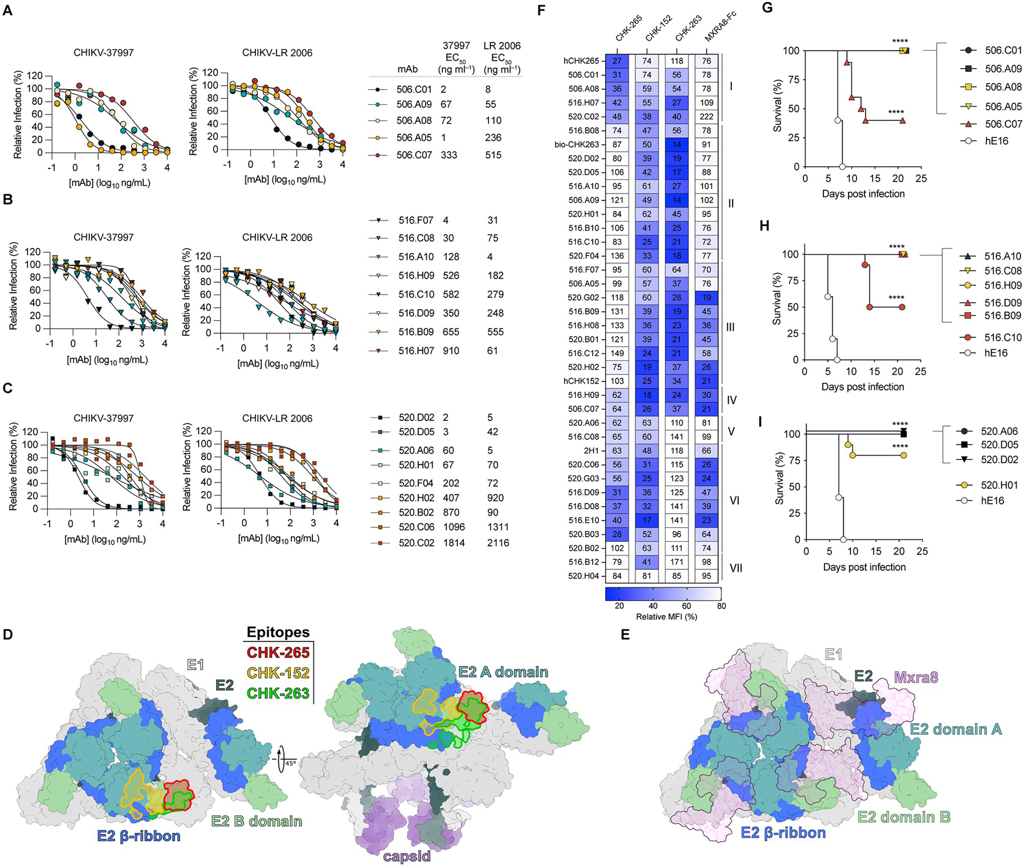
Chikungunya virus (CHIKV) is a mosquito-transmitted alphavirus that causes epidemics of acute and chronic musculoskeletal disease. Here, we analyzed the human B cell response to a CHIKV-like particle-adjuvanted vaccine (PXVX0317) from samples obtained from a phase 2 clinical trial in humans (NCT03483961). Immunization with PXVX0317 induced high levels of neutralizing antibody in serum against CHIKV and circulating antigen-specific B cells up to 6 months after immunization. Monoclonal antibodies (mAbs) generated from peripheral blood B cells of three PXVX0317-vaccinated individuals on day 57 after immunization potently neutralized CHIKV infection, and a subset of these inhibited multiple related arthritogenic alphaviruses. Epitope mapping and cryo-electron microscopy defined two broadly neutralizing mAbs that uniquely bind to the apex of the B domain of the E2 glycoprotein. These results demonstrate the inhibitory breadth and activity of the human B cell response induced by the PXVX0317 vaccine against CHIKV and potentially other related alphaviruses.


Conflict of interest statement

Competing Financial Interests. M.S.D. is a consultant for Inbios, Vir Biotechnology, Senda Biosciences, Moderna, and Immunome. The Diamond laboratory has received unrelated funding support in sponsored research agreements from Moderna, Vir Biotechnology, and Immunome. J.E.C. has served as a consultant for Luna Labs USA, Merck Sharp & Dohme Corporation, and GlaxoSmithKline, is a member of the Scientific Advisory Board of Meissa Vaccines, a former member of the Scientific Advisory Board of Gigagen (Grifols) and is founder of IDBiologics. The laboratory of J.E.C. received unrelated sponsored research agreements from AstraZeneca, Takeda, and IDBiologics during the conduct of the study. K.L.W and L.V. are employees and shareholders of Emergent BioSolutions Inc.

Figures

Figure 1.
Figure 1.. Humoral and B cell response to PXVX00317 Vaccination.
A, Schematic of vaccination regimen and specimen collection. B-C, IgG ELISA (B) against CHIKV VLPs (strain 37997) from serum of immunized subjects from (A) obtained at the indicated time points with interpolated endpoint titers in (c). D-F, EC50 values from FRNT with serum of immunized subjects against CHIKV-37997 (D), CHIKV-LR 2006 (E), and CHIKV RSU1 (F); red circles denote subject 513 who demonstrated pre-existing antibody titers. Data are shown as the mean of 2 experiments performed in technical duplicate. Dotted lines indicate the limit of detection (LOD) of the assay. G, Table of percent of subjects whose Day 57 serum samples demonstrated EC80 or EC50 values by FRNT at a dilution of 1/40 against the indicated arthritogenic alphaviruses. Data are representative of 2 experiments. H-I, Flow cytometry plots (H) of a representative subject showing expression of CD38 and CD71 and binding of VLP to CD19+IgD B cells at the indicated time points following vaccinations, summarized in (I). Bars in (I) represent mean values. ** p < 0.01, ****p < 0.0001 by ANOVA with Sidak’s post-test.
Figure 2.
Figure 2.. Isolation of anti-CHIKV mAbs from vaccine recipients.
A, Merged uniform manifold approximation and projection (UMAP) feature plots of single-cell RNA-seq of 13,764 cells from 3 subjects showing expression of the indicated markers. B, UMAP plots of CD19+ B cells (from subjects 506–3276, 516–6874, and 520–3614) with productive V-D-J contigs showing normalized SAV-TotalSeqC reads corresponding to VLP binding. C, Heatmap of variable heavy chain usage corresponding to each individual from mAbs expressed recombinantly. D, Quantification of mutations in the variable region of the heavy chain from recombinantly expressed mAbs from each subject. E, Heat map of ELISA binding to CHIKV VLP, p62-E1, or E2, and neutralization of CHIKV-LR 2006 by recombinantly expressed mAbs from VLP-binding B cells identified from (A). Intensity of heatmap corresponds to endpoint titer (N = no detectable binding) for ELISA and binned EC50 values for neutralization. Data in (E) are representative of 2 experiments.
Figure 3.
Figure 3.. Characterization of vaccine-elicited mAbs
A-C, Dose-response curves and table of EC50 values of indicated mAbs from subjects 506 (A), 516 (B), and 520 (C) against CHIKV-37997 and CHIKV-LR 2006 by FRNT. Plots in A-C are representative of 4 experiments with EC50 values generated from the mean of all experiments. D, CHIKV asymmetric unit (PDB: 6NK5) with E1 shown in light gray and capsid shown in purple. E2 is colored by domain with B domain in pale green, A domain in teal, and β-ribbon in royal blue, with the remainder of E2 colored dark gray. The footprints of indicated mAbs are highlighted in red (CHK-265), green (CHK-263), and yellow (CHK-152). E, CHIKV asymmetric unit (PDB: 6NK6), colored as in (e). Bound MXRA8 is shown in purple. F, Heatmap of relative binding of each mAb (rows) in the presence of reference mouse mAbs or MXRA8-mouse Fc (columns). Binding to Expi293 cells expressing CHIKV structural proteins (capsid-E3-E2–6k-E1) was assessed by flow cytometry by measuring the mean fluorescence intensity (MFI). Numbers indicate % MFI in the presence of competitor (mAb or MXRA8-mouse Fc) compared to no competitor. G-I, Survival of 4-week-old male C57BL/6J mice treated with 500 μg of anti-IFNAR1 and indicated anti-CHIKV mAb (100 μg, ~5 mg/kg) one day prior to subcutaneous inoculation of 10 FFU of CHIKV-LR 2006. MAbs were isolated from subjects 506 (G), 516 (H), and 520 (I). Data are combined from 2 experiments with n = 10 mice per group. Kaplan-Meier survival curve analysis (log-rank test) with Bonferroni correction compared to isotype control mAb (anti-WNV, hE16); **** (p < 0.0001).
Figure 4.
Figure 4.. Epitope mapping of protective mAbs.
A, Table showing percent relative binding of each mAb (columns) against indicated residues mutated to alanine (rows). The value in each cell correspond to % binding compared to WT and are the mean of 2 experiments. Cells colored in blue are residues also identified by neutralization escape. B, CHIKV E1-E2 trimer (PDB: 6NK5) surface representation, with E1 shown in light gray, E2 B domain in pale green, E2 A domain in teal, E2 β-ribbon in royal blue, and the remainder of E2 colored dark gray. Alanine scanning hits are colored red. A single E1-E2 heterodimer is outlined in magenta. C-F, Ribbon diagrams highlighting alanine scanning hits on E1-E2 heterodimer. Structural proteins are colored as in (B). Antibodies are clustered by site, including those binding B domain sites (apex and flank), both B domain and β-ribbon, or A domain. Alanine scanning mutagenesis loss-of-binding residues shared by all mAbs in each cluster are shown in yellow, with others reducing binding of subsets of mAbs shown in red.
Figure 5.
Figure 5.. Vaccine-induced mAbs protect against related arthritogenic alphaviruses.
A, Heatmap displaying normalized MFI of cell-surface binding of mAbs in Vero cells inoculated with the indicated alphaviruses. Normalized MFI was calculated by dividing geometric MFI of each mAb by geometric MFI of hE16 (isotype control). B, Flow cytometry plots showing binding of indicated mAbs to infected Vero cells. Data in A-B are representative of 3 experiments. C-D, Dose-response curves (C) and mean EC50 values (D) of mAbs against indicated arthritogenic alphaviruses by FRNT. Data are representative (C) or the mean values (D) of 3 experiments. E-F, Viral titers in indicated tissues in 4-week-old C57BL/6 mice administered mAb one day prior to inoculation with 103 FFU of MAYV (E) or RRV (F). Data are pooled from 2 experiments with n = 7–10 mice per group. * (p< 0.05), ** (p < 0.01), *** (p <0.001), **** (p < 0.0001) by Kruskal-Wallis with Dunn’s post-test correction.
Figure 6.
Figure 6.. Cryo-EM reconstructions of broadly-neutralizing vaccine-induced mAbs and CHK-265 bound to CHIKV VLPs.
A-C, CHIKV VLP reconstructions with bound Fab fragments colored green (506.A08) (A), yellow (506.C01) (B), or pink (CHK-265) (C), and structural proteins colored radially. Equatorial cross-sections are shown as round insets. Axes of symmetry are designated in (A) by a pentagon (5-fold; i5), triangles (3-fold; i3), three-pointed stars (quasi-3-fold; q3), and a diamond (2-fold; i2), with axial orientations displayed in the inset. D-F, Asymmetric unit reconstructions with Fabs colored dark green/lime green (506.A08 heavy/light chain), dark gold/yellow (506.C01 heavy/light chain), or crimson/pink (CHK-265 heavy/light chain). CHIKV E1 is shown in white, and capsid is shown in purple. E2 is colored by domain, with the B domain in light green, A domain in teal, and β-ribbon in royal blue, with the remainder of E2 colored dark gray. G, Magnified region from black boxes in D-F and colored as in D-F. A surface representation of E1-E2, with superimposed ribbon diagrams of 506.A08, 506.C01, and CHK-265 Fv fragments. H, Side (left, right) and top (center) views of magnified region from D-F, with mAb footprints outlined in green (506.A08), yellow (506.C01), and red (CHK-265). Residue hits from scanning mutagenesis or neutralization escape are labeled, with impacted mAb(s) indicated by colored stars. I, Alignment of E2 B domains from arthritogenic alphaviruses, including CHIKV-37997, CHIKV-LR 2006, ONNV, GETV, BEBV, RRV, MAYV, and UNAV. Viruses that escape cross-recognition by 506.A08 or 506.C01 are designated by green or yellow diamonds, respectively. Antibody contact residues (determined by PISA solvent exclusion analysis) are delineated below the alignment, with scanning mutagenesis or neutralization escape residues designated by stars. Within the alignment, residues implicated in neutralization escape and correlating with loss of cross-recognition are colored magenta.

References

    1. Suhrbier A, Jaffar-Bandjee M-C, Gasque P, Arthritogenic alphaviruses--an overview. Nat. Rev. Rheumatol. 8, 420–429 (2012). - PubMed
    1. Zacks MA, Paessler S, Encephalitic alphaviruses. Vet. Microbiol. 140, 281–286 (2010). - PMC - PubMed
    1. Schilte C, Staikowsky F, Couderc T, Madec Y, Carpentier F, Kassab S, Albert ML, Lecuit M, Michault A, Chikungunya virus-associated long-term arthralgia: a 36-month prospective longitudinal study. PLoS Negl. Trop. Dis. 7, e2137 (2013). - PMC - PubMed
    1. Burt FJ, Chen W, Miner JJ, Lenschow DJ, Merits A, Schnettler E, Kohl A, Rudd PA, Taylor A, Herrero LJ, Zaid A, Ng LFP, Mahalingam S, Chikungunya virus: an update on the biology and pathogenesis of this emerging pathogen. Lancet Infect. Dis. 17, e107–e117 (2017). - PubMed
    1. Rezza G, Weaver SC, Chikungunya as a paradigm for emerging viral diseases: Evaluating disease impact and hurdles to vaccine development. PLoS Negl. Trop. Dis. 13, e0006919 (2019). - PMC - PubMed

Publication types