Review
. 2023 Sep 15:9:177-199.
doi: 10.1146/annurev-vision-112122-020950. Epub 2023 May 17.

Structure, Function, and Molecular Landscapes of the Aging Retina

Affiliations
Review

Structure, Function, and Molecular Landscapes of the Aging Retina

Jeffrey D Zhu et al. Annu Rev Vis Sci. .

Abstract

Because the central nervous system is largely nonrenewing, neurons and their synapses must be maintained over the lifetime of an individual to ensure circuit function. Age is a dominant risk factor for neural diseases, and declines in nervous system function are a common feature of aging even in the absence of disease. These alterations extend to the visual system and, in particular, to the retina. The retina is a site of clinically relevant age-related alterations but has also proven to be a uniquely approachable system for discovering principles that govern neural aging because it is well mapped, contains diverse neuron types, and is experimentally accessible. In this article, we review the structural and molecular impacts of aging on neurons within the inner and outer retina circuits. We further discuss the contribution of non-neuronal cell types and systems to retinal aging outcomes. Understanding how and why the retina ages is critical to efforts aimed at preventing age-related neural decline and restoring neural function.

Keywords: aging; neuron; retina; synapse.


Figures

Figure 1
Figure 1
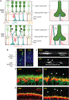
Structural impacts of aging in the retina. Shown is a schematic of retinal neuron organization. In young animals, the outer nuclear layer (ONL) contains rods (dark green) and cones (light green) that form connections in the outer plexiform layer (OPL) with rod bipolar cells (RBPs; ), cone bipolar cells (CBPs; ), and horizontal cells (HCs; red) found in the inner nuclear layer (INL). Also present in the INL are amacrine cells (ACs; blue) that form connections in the inner plexiform layer (IPL). They do so together with bipolar cell axons, which connect with retinal ganglion cell (RGC) dendrites (magenta). RGC cell bodies reside in the ganglion cell layer (GCL). Müller glia (MG; brown) span the length of the outer retina, astrocytes (Astro; orange) reside in the GCL, and microglia (MicroG; yellow) processes are largely restricted to the synaptic layers. Three intraretina vascular layers also interdigitate the GCL, IPL, and OPL. In the aged retina, several of these organizational features are altered. In the outer retina, supporting retinal pigment epithelium (RPE) cells accumulate lipofuscin, and Bruch’s membrane appears to thicken. Rod photoreceptors retract into the ONL, accompanied by misplaced synapses, while cones are structurally less affected. Postsynaptic HC axons and rod bipolar dendrite cells extend neurites into the ONL to maintain contact with retracting rods. These changes are accompanied by synapse misorganization and loss. In the inner retina, the total number of synapses in the IPL decreases, and RGC dendritic arbors decrease in size. Microglia increase in number and become less reactive in injury, while the processes of both Müller glia and astrocytes are attenuated. Vascular function can also decline in both the choroid and the inner retina.
Figure 2
Figure 2
Age-related changes to cell types of the outer retina. (a) Schematic and representative histology images from young (2 months) and (24 months) mice. In the young retina, all rod and cone terminals reside in the outer plexiform layer, where they contact the dendritic terminals of bipolar cells and the axons and dendrites of horizontal cells (HCs), which also remain restricted to this region. With age, photoreceptor density is reduced, and approximately half of rod axons retract their terminals into the outer nuclear layer (black arrowheads), accompanied by HC and rod bipolar spouting (white arrowheads). (b–c) Histological studies of (b) single rod photoreceptors or (c) single HCs labeled with AAV-GFP confirm these alterations, with rod axons retracting (white star), accompanied by sprouting of HC axons (white arrows), but not dendrites. Cones do not undergo similar changes. (d) HC axons (Calbindin, red) and rod bipolar cell dendrites (PKCα, green) sprout far into the outer nuclear layer (white arrowheads). (e) These changes are accompanied by synapse remolding (white arrows; Bassoon, green; Calbindin, red).
Figure 3
Figure 3
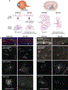
Age-related changes to cell types of the inner retina. (a) Schematic and representative histology images from young (2 months) and (24 months) mice are shown. In the young retina, retinal ganglion cells (RGCs) target distinct lamina in the inner plexiform layer (IPL) and have normal dendrite and axon areas in the retina and brain, respectively. With age, RGC laminar targeting is maintained, but both dendritic and axon arbor area are reduced, with the greatest impact on arbor size in the brain (approximately 30% reduction). (b–c) Representative cross section (upper panel) and whole mount images (lower panels) of age-related alterations to RGC dendrites and axons in young (2 months) and (>2 years) mice are shown. These images highlight that changes occur both (b) in genetically identified RGC subtypes, such as cells that express Junction Adhesion Molecule B (JAM-B), called J-RGCs (see Kim et al. 2008), and (c) in RGC types more generally labeled in the YFPH line in which YFP is driven by elements of the Thy1 promotor (see Feng et al.2000).

References

    1. Aggarwal P, Nag TC, Wadhwa S. 2007. Age-related decrease in rod bipolar cell density of the human retina: an immunohistochemical study. J. Biosci 32:293–98 - PubMed
    1. Alessi DR, Sakamoto K, Bayascas JR. 2006. LKB1-dependent signaling pathways. Annu. Rev. Biochem. 75:137–63 - PubMed
    1. Anderson SR, Roberts JM, Ghena N, Irvin EA, Schwakopf J, et al..2022.Neuronal apoptosis drives remodeling states of microglia and shifts in survival pathway dependence. eLife 11:e76564 - PMC - PubMed
    1. Anderson SR, Roberts JM, Zhang J, Steele MR, Romero CO, et al. 2019. Developmental apoptosis promotes a disease-related gene signature and independence from CSF1R signaling in retinal microglia. Cell Rep. 27:2002–13.e2005 - PMC - PubMed
    1. Apfeld J, O’Connor G, McDonagh T, DiStefano PS, Curtis R. 2004. The AMP-activated protein kinase AAK-2 links energy levels and insulin-like signals to lifespan in C. elegans. Genes Dev. 18:3004–9 - PMC - PubMed

Publication types

LinkOut - more resources