. 2023 May 16;4(5):101045.
doi: 10.1016/j.xcrm.2023.101045.

Molecular signatures of post-traumatic stress disorder in war-zone-exposed veteran and active-duty soldiers

Affiliations

Molecular signatures of post-traumatic stress disorder in war-zone-exposed veteran and active-duty soldiers

Seid Muhie et al. Cell Rep Med. .

Abstract

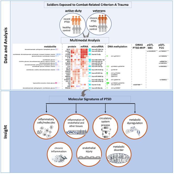
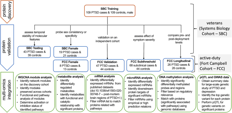
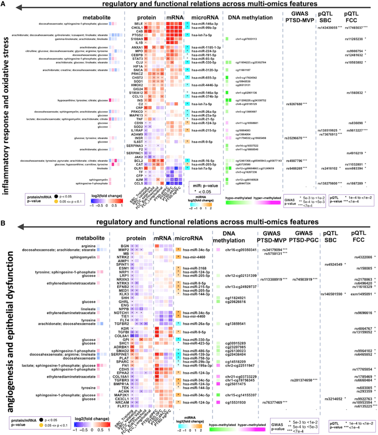
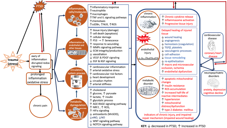
Post-traumatic stress disorder (PTSD) is a multisystem syndrome. Integration of systems-level multi-modal datasets can provide a molecular understanding of PTSD. Proteomic, metabolomic, and epigenomic assays are conducted on blood samples of two cohorts of well-characterized PTSD cases and controls: 340 veterans and 180 active-duty soldiers. All participants had been deployed to Iraq and/or Afghanistan and exposed to military-service-related criterion A trauma. Molecular signatures are identified from a discovery cohort of 218 veterans (109/109 PTSD+/-). Identified molecular signatures are tested in 122 separate veterans (62/60 PTSD+/-) and in 180 active-duty soldiers (PTSD+/-). Molecular profiles are computationally integrated with upstream regulators (genetic/methylation/microRNAs) and functional units (mRNAs/proteins/metabolites). Reproducible molecular features of PTSD are identified, including activated inflammation, oxidative stress, metabolic dysregulation, and impaired angiogenesis. These processes may play a role in psychiatric and physical comorbidities, including impaired repair/wound healing mechanisms and cardiovascular, metabolic, and psychiatric diseases.

Keywords: active duty; angiogenesis; inflammatory response; metabolic dysregulation; molecular signature; multi-omics; oxidative stress; post-traumatic stress disorder; veterans; wound healing.


Conflict of interest statement

Declaration of interests The authors declare no competing interests

Figures

None
Graphical abstract
Figure 1
Figure 1
Overall workflow of the study Identifying, validating, and characterizing of PTSD-associated proteins and integration with multi-modal molecular features from blood samples of veterans and active-duty military participants, published postmortem brain regions, and summary statistics from publicly available genome-wide association studies. PTSD cases and trauma-exposed healthy controls composed of two well-characterized cohorts: Systems Biology Consortium (SBC: 340 veterans) and Fort Campbell Cohort (FCC: 180 active-duty service members). All participants had been deployed to Iraq and/or Afghanistan. The active-duty cohort included blood biomarkers and clinical features assessed longitudinally before and after deployment. Male veterans with chronic PTSD (CAPS scores ≥ 40; ≥3 months duration) and matched controls (CAPS total score < 20) were recruited into training and testing cohorts. A smaller, case-control female veteran cohort with chronic PTSD was recruited with the same inclusion criteria. Active-duty males with recent PTSD (PCL ≥ 38; 3 days before or 90–180 days post-deployment), active-duty males with subthreshold recent PTSD (PCL ≥ 22 to < 38; 3 days before or 90–180 post-deployment), and active-duty females with recent PTSD; all cohorts included matched controls (PCL < 22). Recent onset PTSD case-controls (n = 26) were a longitudinal male active-duty cohort with recent PTSD (n = 26 controls: PCL < 22 at 2 weeks pre-deployment; n = 26 cases: PCL ≥ 31 at 3 days before or 90–180 days post-deployment). The FCC Validation and FCC Subthreshold groups shared the same controls. Comparability of molecular datasets across cohorts was verified by quality control output graphs presented in Figures S1 and S2. CAPS: Clinician-Administered PTSD Scale; PCL: PTSD Checklist.
Figure 2
Figure 2
Modular networks and enriched pathways across cohorts (A) Identification of protein co-expression modules associated with PTSD by weighted gene co-expression network analysis (WGCNA). Module identification by hierarchical clustering tree (dendrogram) of the consensus network comprising 1,305 proteins where branches of the dendrogram grouped together densely interconnected, highly co-expressed proteins. Modules were identified in the SBC Training group (109/109 PTSD+/−), shown in the first band underneath the tree. Colors represent each modular network. Subsequent bands indicate modules in the SBC Testing (43/39 PTSD+/−), FCC Validation (47/44 PTSD+/−), and FCC Subthreshold (68/44 PTSD subclinical/controls) groups. (B) Module preservation identified six highly preserved modules (preservation Z score > 10; ≥30 proteins per module). Four modules (turquoise, yellow, blue, and red) were significantly correlated (p < 0.01) with PTSD across the SBC Testing, FCC Validation, and FCC Subthreshold (Figure S3). (C) Biological processes and pathways identified using hypergeometric enrichment filter at q < 0.05, Bonferroni correction (family-wise error rate) followed by pathway activation analyses. Ranking is based on pathway enrichment significance. (D) Pathway or process significantly associated (FDR [false discovery rate]-corrected) with differentially altered proteins (PTSD cases vs. controls) in the SBC Training, SBC Testing, FCC Validation, FCC Longitudinal, and FCC Subthreshold. (E) Significantly activated or inhibited pathways that were correlated with CAPS total current (SBC cohort) and PCL scores (FCC cohort). Top panel, positive correlation (red gradient); bottom panel, negative correlation (blue gradient). (F and G) Pathways and biological processes significantly associated with member proteins of the methylation-protein consensus modules (RP: receptor protein; arrows indicate Gene Ontology (GO) or pathway hierarchy from stem to leaves; background colors show the different classes of pathways). Bubble plots denote q values (red, activated; blue, inhibited), where size corresponds with number of proteins. Complete data are in Figure S4. CAPS: Clinically Administered PTSD Scale; CAPSTOT_Curr: CAPS current total; CAPSTOT_lt: CAPS lifetime total; CAPSB: CAPS criterion B (re-experiencing); CAPSC: criterion C (avoidance of trauma reminders); CAPSD: CAPS criterion D (negative cognitions and affect).
Figure 3
Figure 3
Correlations among significantly inhibited or activated pathways and PTSD diagnostic variables (A) Significant pathways negatively correlated with CAPS total current (SBC cohort) or PCL scores (FCC cohort). (B) Significant pathways positively correlated with CAPS total current (SBC cohort) or PCL scores (FCC cohort). (C) Correlations between significantly inhibited or activated pathways with wound healing in SBC Training (109/109 PTSD+/−), SBC Testing (43/39 PTSD+/−), and FCC Validating (47/44 PTSD+/−) cohorts. Correlations between wound healing and pathways associated with vasculature (top), inflammation/oxidative stress (middle), and metabolic disorder/obesity (bottom) were evaluated in the SBC Training, SBC Testing, and FCC Validation cohorts. PCL, PTSD Checklist, CAPSTOT_cur: the total current score for Clinically Administered PTSD Scale.
Figure 4
Figure 4
Integrated multi-omics showing regulatory and functional relations (horizontally from right to left) across genetic variants, epigenetic marks, microRNAs, mRNAs proteins, and metabolites (A and B) Differentially expressed proteins (DEPs) that were persistent across PTSD cohorts and associated with (A) activated inflammatory response or oxidative stress, (B) impaired angiogenesis, epithelial dysfunction, or cardiovascular function were integrated with multi-omics datasets and compared across SBC Training (109/109 PTSD+/−), SBC Testing (43/39 PTSD+/−), FCC Validation (47/44 PTSD+/−), FCC Longitudinal (26/26 PTSD+/−), and FCC Subthreshold (68/44 PTSD subclinical/controls) cohorts. Vertical lanes of the protein heatmap correspond to the fold changes of each protein from each group of cohorts (as shown by the labels). SBC: Systems Biology Consortium (veteran cohort), FCC: Fort Campbell (active-duty) Cohort; for brain regions (postmortem mRNA data): dIPFC, dorsolateral pre-frontal cortex (PFC); ACC, anterior cingulate cortex (ACC); dACC, dorsal ACC; sgPFC, subgenual PFC; OFC, orbito-frontal cortex.
Figure 5
Figure 5
Summary of biological processes and pathways correlated with PTSD clinicals identified in integrated multi-omics analyses across cohorts Multi-omics analyses identified inhibition and activation of specific components of pathways associated with impaired wound healing and comorbidities indicative of chronic inflammation, endothelial injury, and metabolic disorders. Disrupted and prolonged inflammation and redox signaling leading to inflammation and injuries of endothelium and other tissues, metabolic dysregulation, and circulatory system dysfunction. The associated physiological dysregulations correspond with long-term sequelae of PTSD, including cardiovascular disease, T2DM and neuropsychiatric disorders (anxiety, depression, and cognitive decline). Although our study evaluated peripheral markers, the processes identified may either reflect system-wide (including CNS) perturbations or else may lead systemic disruptions related to PTSD pathology. Up arrows indicate upregulation/activation in PTSD; down arrows indicate downregulation/inhibition.

References

    1. PTSD N.C.f. How common is PTSD? 2022. https://www.ptsd.va.gov/understand/common/common_veterans.asp
    1. Mellon S.H., Bersani F.S., Lindqvist D., Hammamieh R., Donohue D., Dean K., Jett M., Yehuda R., Flory J., Reus V.I., et al. Metabolomic analysis of male combat veterans with post traumatic stress disorder. PLoS One. 2019;14 doi: 10.1371/journal.pone.0213839. - DOI - PMC - PubMed
    1. Somvanshi P.R., Mellon S.H., Flory J.D., Abu-Amara D., PTSD Systems Biology Consortium; Wolkowitz O.M., Yehuda R., Jett M., Hood L., Marmar C., Doyle F.J., 3rd. Mechanistic inferences on metabolic dysfunction in posttraumatic stress disorder from an integrated model and multiomic analysis: role of glucocorticoid receptor sensitivity. Am. J. Physiol. Endocrinol. Metab. 2019;317:E879–E898. doi: 10.1152/ajpendo.00065.2019. - DOI - PMC - PubMed
    1. Yang R., Gautam A., Getnet D., Daigle B.J., Miller S., Misganaw B., Dean K.R., Kumar R., Muhie S., Wang K., et al. Epigenetic biotypes of post-traumatic stress disorder in war-zone exposed veteran and active duty males. Mol. Psychiatry. 2021;26:4300–4314. doi: 10.1038/s41380-020-00966-2. - DOI - PMC - PubMed
    1. Katrinli S., Stevens J., Wani A.H., Lori A., Kilaru V., van Rooij S.J.H., Hinrichs R., Powers A., Gillespie C.F., Michopoulos V., et al. Evaluating the impact of trauma and PTSD on epigenetic prediction of lifespan and neural integrity. Neuropsychopharmacology. 2020;45:1609–1616. doi: 10.1038/s41386-020-0700-5. - DOI - PMC - PubMed

Publication types

MeSH terms