. 2023 Jun 14;31(6):937-948.e4.
doi: 10.1016/j.chom.2023.05.001. Epub 2023 May 5.

Complement-dependent mpox-virus-neutralizing antibodies in infected and vaccinated individuals

Affiliations

Complement-dependent mpox-virus-neutralizing antibodies in infected and vaccinated individuals

Mathieu Hubert et al. Cell Host Microbe. .

Abstract

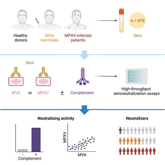
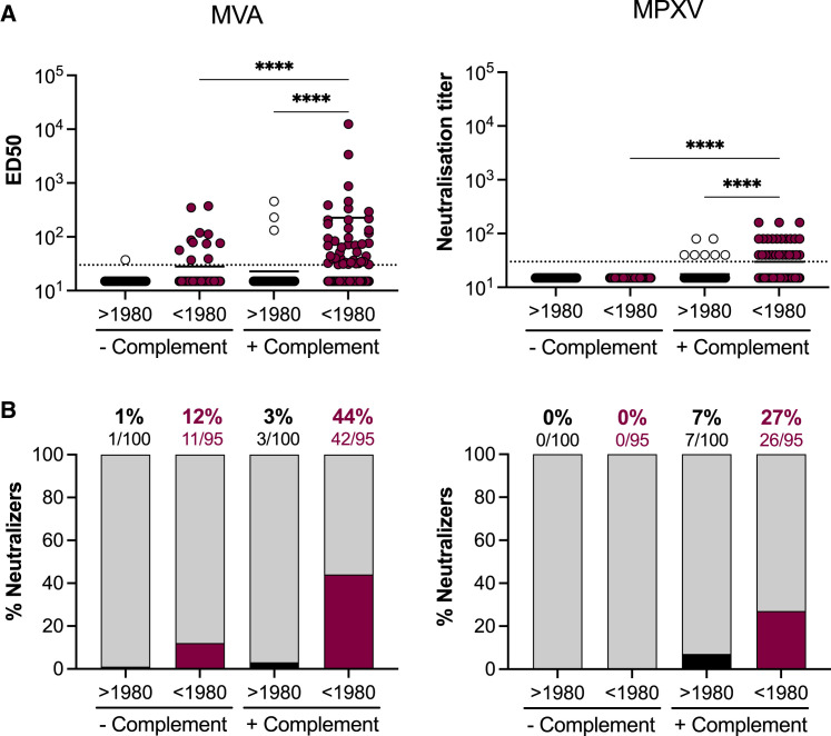
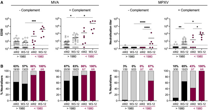
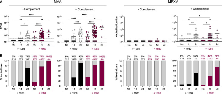
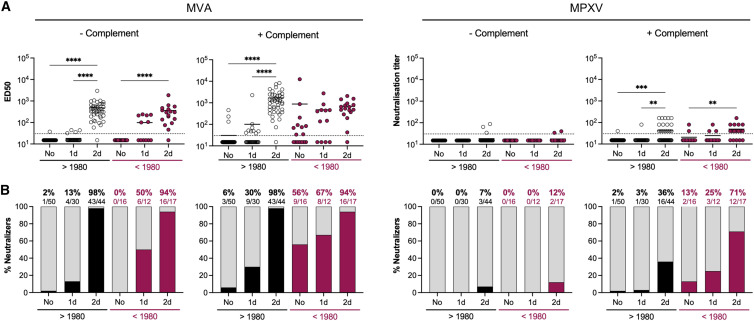
Mpox virus (MPXV) caused a multi-country outbreak in non-endemic areas in 2022. Following historic success of smallpox vaccination with vaccinia virus (VACV)-based vaccines, the third generation modified vaccinia Ankara (MVA)-based vaccine was used as prophylaxis for MPXV, but its effectiveness remains poorly characterized. Here, we applied two assays to quantify neutralizing antibodies (NAbs) in sera from control, MPXV-infected, or MVA-vaccinated individuals. Various levels of MVA NAbs were detected after infection, historic smallpox, or recent MVA vaccination. MPXV was minimally sensitive to neutralization. However, addition of complement enhanced detection of responsive individuals and NAb levels. Anti-MVA and -MPXV NAbs were observed in 94% and 82% of infected individuals, respectively, and 92% and 56% of MVA vaccinees, respectively. NAb titers were higher in individuals born before 1980, highlighting the impact of historic smallpox vaccination on humoral immunity. Altogether, our results indicate that MPXV neutralization is complement dependent and uncover mechanisms underlying vaccine effectiveness.

Keywords: complement; hybrid immunity; mpox virus; neutralizing antibodies; smallpox vaccination.


Conflict of interest statement

Declaration of interests The authors declare no competing interests.

Figures

None
Graphical abstract
Figure 1
Figure 1
The MVA-GFP seroneutralization assay (A) A Modified vaccinia virus Ankara expressing GFP (MVA-GFP) was mixed to serial dilutions of sera to be tested in the absence or the presence of 10% guinea pig serum as a source of complement. After 30 min to 2 h, the mixture was added onto Vero E6 target cells. After 20 h, GFP+ infected cells were quantified by high-content confocal microscopy and the percentage of neutralization was calculated. (B) Vero E6 cells were exposed to serially diluted MVA-GFP. After 20 h, the GFP+ area was quantified. Left: representative images of MVA-GFP-infected cells. The numbers indicate the dilution factor of the viral inoculum. Scale bars: 500 μm. Right: standard curve of the GFP signal. (C) Examples of MVA-GFP neutralization by 4-fold serial dilutions of two plasma from the same MPXV-infected patient at 2 and 56 days after the onset of symptoms. Left: examples of micrographs. The numbers indicate the percentage of neutralization by considering the no serum and non-infected conditions as 0% and 100% neutralization, respectively. Scale bars: 500 μm. Right: activity of the non-neutralizing (red) and neutralizing plasma (gray). The median effective dose (ED50) represents the plasma dilution reducing infection by 50%. (D) Neutralization of a serum from an IMVANEX vaccinee tested with several MVA-GFP inocula, in the absence (left) or the presence (right panel) of complement. Representative graphs are shown. (E) Example of neutralization of MVA-GFP (dilution factor: 2 × 104) by a serum from an IMVANEX vaccinee.
Figure 2
Figure 2
The MPXV seroneutralization assay (A) Mpox virus (MPXV) was mixed to serial dilutions of sera to be tested, in the absence or the presence of 10% guinea pig serum as a source of complement. After 30 min to 2 h, the mixture was added to U2OS target cells. After 48 h, the cells were fixed. MPXV infection was monitored by immunofluorescence using a rabbit polyclonal anti-VACV antibody. The percentage of neutralization was calculated. (B) U2OS cells were exposed to serially diluted MPXV. After 48 h, cells were fixed and stained. Left: representative images of MPXV-infected cells. The numbers indicate the dilution factor of the viral inoculum. Scale bars: 500 μm. Right: standard curve of the signal. (C) Examples of MPXV neutralization by 2-fold serial dilutions of a non-neutralizing serum (uninfected individual) and a neutralizing serum (MPXV-infected patient). The white symbols show the presence (+) or absence (−) of neutralization. Scale bars: 500 μm. The neutralizing titer was calculated by selecting the highest dilution in which neutralization was observed.
Figure 3
Figure 3
Basal levels of neutralizing antibodies in uninfected individuals (A) Seroneutralization of MVA-GFP (left) and MPXV (right) by sera from uninfected individuals in the absence and the presence of 10% guinea pig serum as a source of complement. Sera from individuals born before and after 1980 were analyzed. See also Table S1. The dotted lines represent the limit of detection (LOD). Each dot represents an individual and data are mean of 2–6 independent experiments. Bars indicate mean values. Statistical analysis: Kruskal-Wallis tests with Dunn’s multiple comparisons correction (∗∗∗∗p < 0.0001). (B) The proportion of neutralizers was estimated as the percentage of individuals exhibiting a neutralizing activity > LOD.
Figure 4
Figure 4
Neutralizing antibodies induced by MPXV infection (A) Seroneutralization of MVA-GFP (left) and MPXV (right) by sera from MPXV-infected patients in the absence and the presence of 10% guinea pig serum as a source of complement. Sera from individuals born before and after 1980 were analyzed. The weeks of sample collection are shown (≤W2: samples collected during the two first weeks after onset of symptom; W3–12: samples collected between the 3rd and 12th weeks). See also Table S2. The dotted lines represent the limit of detection (LOD). Each dot represents an individual and data are mean of two to six independent experiments. Bars indicate mean values. Statistical analysis: Kruskal-Wallis tests with Dunn’s multiple comparisons correction (p < 0.03; ∗∗p < 0.002; ∗∗∗p < 0.0002; ∗∗∗∗p < 0.0001). (B) The proportion of neutralizers was estimated as the percentage of individuals exhibiting a neutralizing activity > LOD.
Figure 5
Figure 5
Neutralizing antibodies induced by IMVANEX vaccination (A) Seroneutralization of MVA-GFP (left) and MPXV (right) by sera from IMVANEX vaccinees in the absence and the presence of 10% guinea pig serum as a source of complement. Sera from individuals born before and after 1980 were analyzed. The vaccination status at the time of sample collection is indicated (no: samples collected before vaccination; 1d: samples collected after the 1st dose; 2d: samples collected after the 2nd dose). See also Table S3. The dotted lines represent the limit of detection (LOD). Each dot represents an individual, and data are mean of two to six independent experiments. Bars indicate mean values. Statistical analysis: Kruskal-Wallis tests with Dunn’s multiple comparisons correction (p < 0.03; ∗∗p < 0.002; ∗∗∗p < 0.0002; ∗∗∗∗p < 0.0001). (B) The proportion of neutralizers was estimated as the percentage of individuals exhibiting a neutralizing activity > LOD.
Figure 6
Figure 6
Neutralizing antibodies induced by an experimental MVA-HIV vaccine (A) Seroneutralization of MVA-GFP (left) and MPXV (right) by sera from IMVANEX vaccinees in the absence and the presence of 10% guinea pig serum as a source of complement. Sera from individuals born before and after 1980 were analyzed. The vaccination status at the time of sample collection is indicated. (No: samples collected before vaccination; 1d: samples collected after the 1st dose; 2d: samples collected after the 2nd dose.) See also Table S4. The dotted lines represent the limit of detection (LOD). Each dot represents an individual and data are mean of two to six independent experiments. Bars indicate mean values. Statistical analysis: Kruskal-Wallis tests with Dunn’s multiple comparisons correction (p < 0.03; ∗∗p < 0.002; ∗∗∗p < 0.0002; ∗∗∗∗p < 0.0001). (B) The proportion of neutralizers was estimated as the percentage of individuals exhibiting a neutralizing activity > LOD.

References

    1. Gessain A., Nakoune E., Yazdanpanah Y. Monkeypox. N. Engl. J. Med. 2022;387:1783–1793. doi: 10.1056/NEJMra2208860. - DOI - PubMed
    1. Wang L., Shang J., Weng S., Aliyari S.R., Ji C., Cheng G., Wu A. Genomic annotation and molecular evolution of monkeypox virus outbreak in 2022. J. Med. Virol. 2023;95:e28036. doi: 10.1002/jmv.28036. - DOI - PMC - PubMed
    1. Ligon B.L. Monkeypox: a review of the history and emergence in the western hemisphere. Semin. Pediatr. Infect. Dis. 2004;15:280–287. doi: 10.1053/j.spid.2004.09.001. - DOI - PMC - PubMed
    1. Adler H., Gould S., Hine P., Snell L.B., Wong W., Houlihan C.F., Osborne J.C., Rampling T., Beadsworth M.B., Duncan C.J., et al. Clinical features and management of human monkeypox: a retrospective observational study in the UK. Lancet Infect. Dis. 2022;22:1153–1162. doi: 10.1016/S1473-3099(22)00228-6. - DOI - PMC - PubMed
    1. Erez N., Achdout H., Milrot E., Schwartz Y., Wiener-Well Y., Paran N., Politi B., Tamir H., Israely T., Weiss S., et al. Diagnosis of imported monkeypox, Israel, 2018. Emerg. Infect. Dis. 2019;25:980–983. doi: 10.3201/eid2505.190076. - DOI - PMC - PubMed