. 2023 Oct;44(10):1935-1947.
doi: 10.1038/s41401-023-01100-y. Epub 2023 May 17.

A novel small-molecular CCR5 antagonist promotes neural repair after stroke

Affiliations

A novel small-molecular CCR5 antagonist promotes neural repair after stroke

Qing-Lin Wu et al. Acta Pharmacol Sin. 2023 Oct.

Abstract

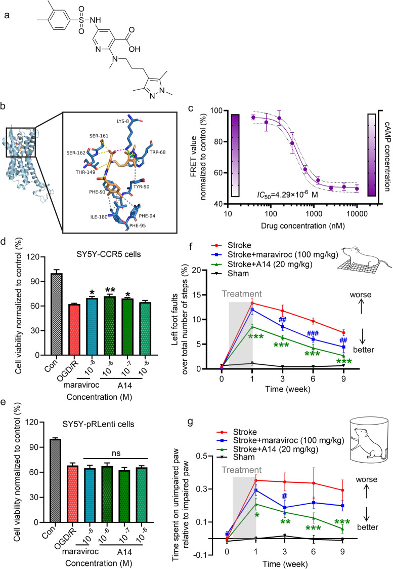
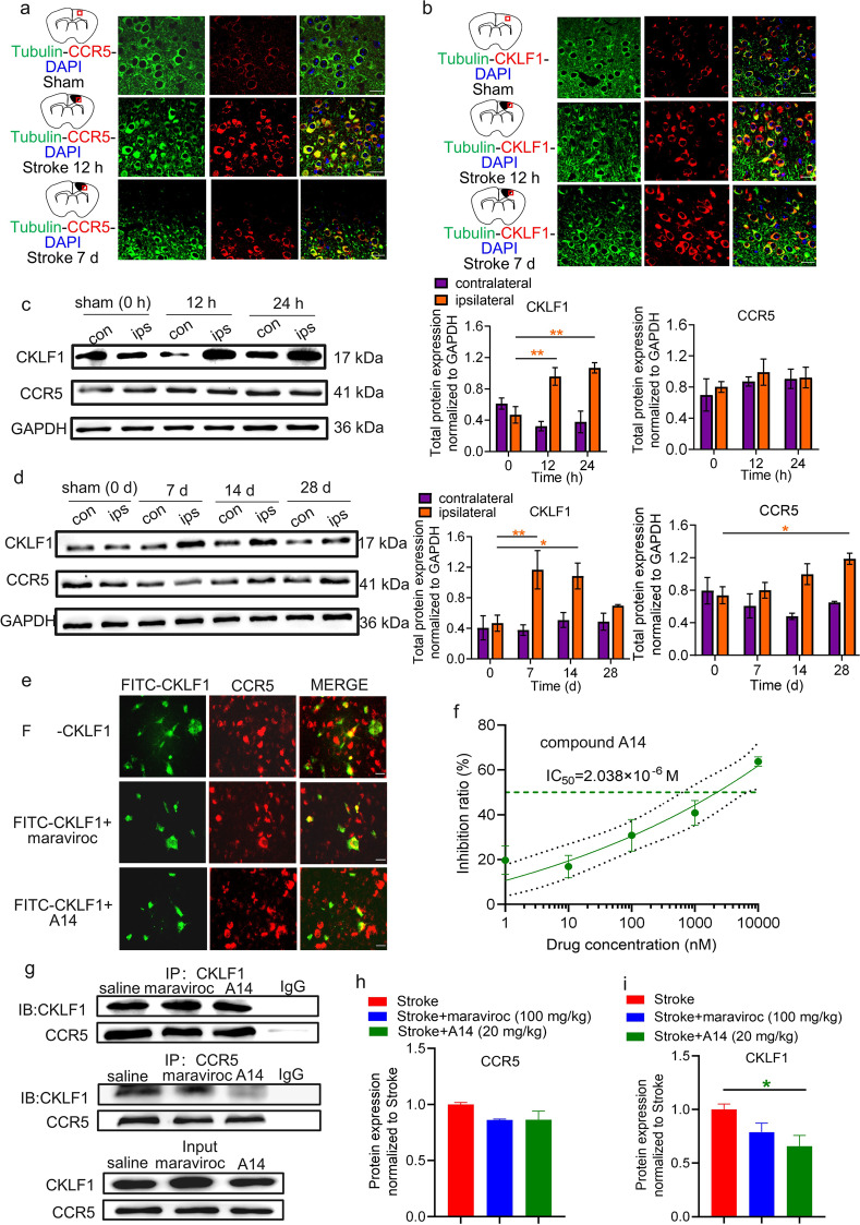
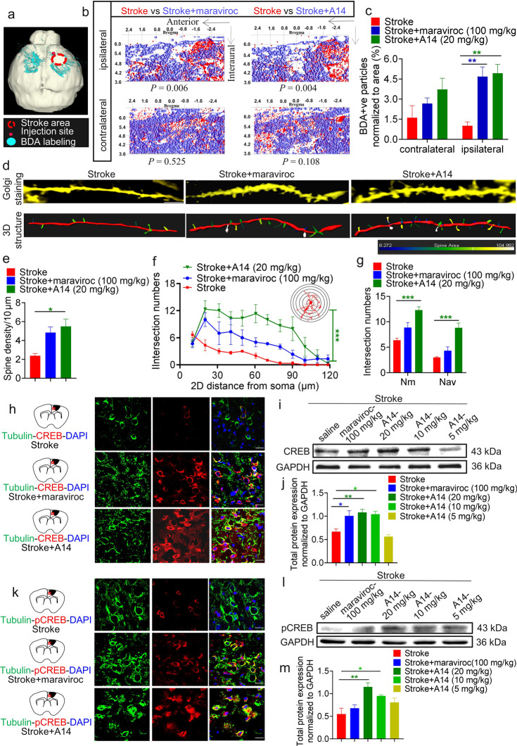
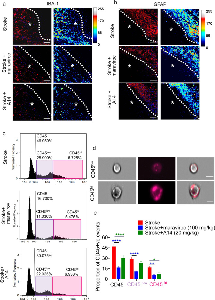
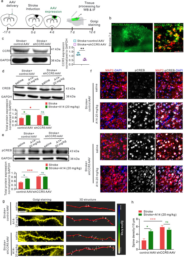
Chemokine receptor 5 (CCR5) is one of the main co-receptors of HIV-1, and has been found to be a potential therapeutic target for stroke. Maraviroc is a classic CCR5 antagonist, which is undergoing clinical trials against stroke. As maraviroc shows poor blood-brain barrier (BBB) permeability, it is of interest to find novel CCR5 antagonists suitable for neurological medication. In this study we characterized the therapeutic potential of a novel CCR5 antagonist A14 in treating ischemic stroke mice. A14 was discovered in screening millions compounds in the Chemdiv library based on the molecular docking diagram of CCR5 and maraviroc. We found that A14 dose-dependently inhibited the CCR5 activity with an IC50 value of 4.29 μM. Pharmacodynamic studies showed that A14 treatment exerted protective effects against neuronal ischemic injury both in vitro and vivo. In a SH-SY5Y cell line overexpressing CCR5, A14 (0.1, 1 μM) significantly alleviated OGD/R-induced cell injury. We found that the expression of CCR5 and its ligand CKLF1 was significantly upregulated during both acute and recovery period in focal cortical stroke mice; oral administration of A14 (20 mg·kg-1·d-1, for 1 week) produced sustained protective effect against motor impairment. A14 treatment had earlier onset time, lower onset dosage and much better BBB permeability compared to maraviroc. MRI analysis also showed that A14 treatment significantly reduced the infarction volume after 1 week of treatment. We further revealed that A14 treatment blocked the protein-protein interaction between CCR5 and CKLF1, increasing the activity of CREB signaling pathway in neurons, thereby improving axonal sprouting and synaptic density after stroke. In addition, A14 treatment remarkably inhibited the reactive proliferation of glial cells after stroke and reduced the infiltration of peripheral immune cells. These results demonstrate that A14 is a promising novel CCR5 antagonist for promoting neuronal repair after ischemic stroke. A14 blocked the protein-protein interaction between CKLF1 and CCR5 after stroke by binding with CCR5 stably, improved the infarct area and promoted motor recovery through reversing the CREB/pCREB signaling which was inhibited by activated CCR5 Gαi pathway, and benefited to the dendritic spines and axons sprouting.

Keywords: CCR5 antagonist; CKLF1; CREB signaling; chemokine receptor 5; ischemic stroke; neural repair.


Conflict of interest statement

The authors declare no competing interests.

Figures

None
A14 blocked the protein-protein interaction between CKLF1 and CCR5 after stroke by binding with CCR5 stably, improved the infarct area and promoted motor recovery through reversing the CREB/pCREB signaling which was inhibited by activated CCR5 Gαi pathway, and benefited to the dendritic spines and axons sprouting.
Fig. 1
Fig. 1. A14, an antagonist of CCR5, produced early and sustained motor recovery effects in a stroke model.
a The chemical structure of A14. b The binding mode obtained based on the last frame of Pose2 in the kinetic simulation process, overall view (left) and local view (right). The green stick represents the small molecule (A14), and the light blue cartoon is the protein (CCR5). The yellow dotted line represents hydrogen bonding, the gray line represents hydrophobicity, the wine red dotted line represents salt bridges, and the green dotted line represents cation-pi conjugation or pi-pi conjugation. c A14 blocked CCR5 and increased cAMP concentration in a dose-dependent manner. The IC50 was calculated by nonlinear fit. The 95% confidence limits (Cl) were 2.625 μM to 8.459 μM. d A14 protected against OGD/R-induced neural impairment in a dose-dependent manner. Data are expressed as the mean ± SEM, *P < 0.05, **P < 0.01 vs. OGD/R. The experiment was repeated three times, each time set as n = 3 of each group. e A14 had no protective effect on control cell lines against OGD/R-induced neural impairment. Data are expressed as the mean ± SEM. The experiment was repeated three times, each time set as n = 3 of each group. f, g Oral administration of A14 at a dosage of 20 mg/kg for 1 week produced long-term neural repair. A14 promoted motor recovery after 1 week compared with stroke only (*P < 0.05, **P < 0.01, ***P < 0.001 vs. Stroke). Maraviroc produced motor recovery after 3 weeks compared with stroke (#P < 0.05, ##P < 0.01, ###P < 0.001 vs. Stroke) in the cylinder test (g). n = 9 mice for each group.
Fig. 2
Fig. 2. A14 blocked the interaction between CCR5 and CKLF1.
a, b The expression of CKLF1-CCR5 showed spatiotemporal differences post stroke. Representative images of double-staining immunofluorescence of neurons with CCR5 (a) and neurons with CKLF1 (b) in mice at 12 h and 7 d after stroke. Scale bars, 20 μm. Representative blots and densitometry data of CKLF1 and CCR5 expression in the cortex of ipsilateral and contralateral brain hemispheres of mice during the acute stage (c) and recovery stage (d) after stroke. n = 3 mice per group. Each experiment represented by one image was repeated three times. Data are expressed as the mean ± SEM, *P < 0.05, **P < 0.01 vs. Sham. e, f A14 blocked the binding between CKLF1 and CCR5. Fluorescence was monitored by fluorescence microscopy when FITC-CKLF1 was combined with SY5Y-CCR5 cells (e). Scale bars, 20 μm. Inhibition of CCR5 with A14 reversed fluorescence release when FITC-CKLF1 was combined with SY5Y-CCR5 cells. Data on inhibition were calculated by 1 − (Fcompound − Fcompound control)/ (FCKLF1 − FCKLF1 control). The IC50 was calculated by nonlinear fit, and the 95% CI was 0.8733 μM to 6.073 μM (f). The experiment was repeated three times, each time set as n = 3 of each group. gi A14 (20 mg/kg) decreased the interaction between CKLF1 and CCR5 in vivo. Representative blots of the Co-IP assay for CKLF1 in CCR5 and CCR5 in CKLF1 in the ischemic cortex of mice at 7 d post stroke (g). Quantitative analysis of CCR5 co-immunoprecipitated with CKLF1 and CKLF1 co-immunoprecipitated with CCR5 (h, i). n = 3 mice for each group. Data are expressed as the mean ± SEM, *P < 0.05 vs. Stroke.
Fig. 3
Fig. 3. Axon sprouting and dendrite spine density were enhanced by A14, accompanied by an improved CREB signaling pathway.
ac A14 (20 mg/kg) induced axonal sprouting in the ipsilateral cortex after stroke. Schematic diagram of BDA labeling axons after 11 weeks of stroke (a). Axes represent approximate stereotactic coordinates anterior and interaural to bregma (b). Blue represents different treatment groups (maraviroc and A14), red represents the stroke only group. The images are the mapping of BDA-positive points in different lateral positions of the ipsilateral cortex and contralateral cortex in the same plane. n = 3 mice for each group. Data are shown as the mean ± SEM. **P < 0.01 vs. Stroke. dg A14 (20 mg/kg) increased dendritic spines after stroke. Representative Golgi staining and 3D structure of dendrites in the cortical peri-infract area (d). Scale bar, 4 μm. A14 increased dendritic spine density after 9 weeks compared with stroke only (e). n = 3 mice for each group. Data are expressed as the mean ± SEM, *P < 0.05 vs. Stroke. A14 (20 mg/kg) produced neuronal complexity (f, g). Neurons from three mice per group were analyzed by Sholl analysis, ***P < 0.001 vs. Stroke. hj Representative photomicrographs of double-staining immunofluorescence of neurons with CREB in mice at d 7 after stroke (h). Scale bars, 20 μm. Representative blots of CREB expression in the ischemic cortex of mice at d 7 post stroke (i, j). n = 3 mice for each group. Data are expressed as the mean ± SEM, *P < 0.05, **P < 0.01 vs. Stroke. km Representative photomicrographs of double-staining immunofluorescence of neurons with pCREB in mice at d 7 after stroke (k). Scale bars, 20 μm. Representative blots of pCREB expression in the ischemic cortex of mice at d 7 post stroke (l, m). n = 3 mice for each group. Data are expressed as the mean ± SEM, *P < 0.05, **P < 0.01 vs. Stroke.
Fig. 4
Fig. 4. A14 reduced neuroglial reactivity and dampened post-stroke lymphocyte recruitment.
a, b Images represent differential immunoreactivity for the astrocytic marker GFAP and the microglial marker IBA-1 in different treatment conditions at d 7 after stroke. The dotted line denotes the infarct border, and the asterisk denotes the infarct. A14 (20 mg/kg) improved microglial infiltration (a) and astrocyte proliferation (b) in the peripheral area of cerebral infarction after cerebral ischemia. Scale bars, 100 μm. ce Representative histograms (c) and images (d) from FCM show events gated for CD45, CD45low (microglia), and CD45high (peripheral lymphocytes) from stroke animals treated with A14 (20 mg/kg) or maraviroc (100 mg/kg) at d 7 post stroke. Scale bars, 7 μm. The proportion of CD45 + ve cells is quantified in e. n = 4 for each group. Data are mean ± SEM, *P < 0.05, **P < 0.01, ****P < 0.0001 vs. Stroke.
Fig. 5
Fig. 5. A14 treatment for one week reduced infarct volume and promoted axonal sprouting in the ipsilateral cortex after stroke.
ad Treatment with 20 mg/kg A14 decreased the infarct area (a, b) and infarct volume (c, d) after 1 week compared with stroke only. Infarct volume was calculated by (A1 + A2.. + An)t, where A = lesion area from each tangential section collected from the entire flattened cortical tissue and t means thickness of each section = 0.5 mm. n = 5 mice for each group. Data are mean ± SEM, *P < 0.05 vs. Stroke.
Fig. 6
Fig. 6. A14 promoted neural repair after stroke in vivo by targeting CCR5.
a Schedule of CCR5 knockdown experiments. b Images represent shCCR5 AAV in the cortex at d 7 of stroke. Viral expression (EGFP, green) was co-localized with neurons (MAP2 + ve, red). Scale bars, 50 μm. c Representative blot and quantification of CCR5 expression in the injured cortex from mice treated with control AAV or shCCR5 AAV. n = 3 mice for each group. Data are mean ± SEM, **P < 0.01 vs. Stroke+control AAV. d, e Representative blot and quantification of CREB (d) and pCREB (e) expression in the cortex. n = 3 mice for each group. Data are mean ± SEM, *P < 0.05, ***P < 0.001. f Representative images of double-staining immunofluorescence of neurons (MAP2 + ve, red) with pCREB (gray, excited at 647 nm) in mice at d 7 after stroke. Scale bars, 10 μm. g Representative Golgi staining and 3D structure of dendrites in the cortical peri-infarct area. Scale bar, 10 μm. h A14 (20 mg/kg) and CCR5 knockdown increased dendritic spine density at 12 days after stroke compared with Stroke+control AAV, A14 (20 mg/kg) had no significance compared with Stroke+shCCR5, *P < 0.05, ***P < 0.001 vs. Stroke+control AAV. n = 5 mice for each group. Data are expressed as the mean ± SEM.

References

    1. Collaborators GBDS. Global, regional, and national burden of stroke and its risk factors, 1990-2019: a systematic analysis for the Global Burden of Disease Study 2019. Lancet Neurol. 2021;20:795–820. doi: 10.1016/S1474-4422(21)00252-0. - DOI - PMC - PubMed
    1. Collaborators GBDCoD. Global, regional, and national age-sex-specific mortality for 282 causes of death in 195 countries and territories, 1980-2017: a systematic analysis for the Global Burden of Disease Study 2017. Lancet. 2018;392:1736–88. doi: 10.1016/S0140-6736(18)32203-7. - DOI - PMC - PubMed
    1. Lin DJ, Finklestein SP, Cramer SC. New directions in treatments targeting stroke recovery. Stroke. 2018;49:3107–14. doi: 10.1161/STROKEAHA.118.021359. - DOI - PMC - PubMed
    1. Joy MT, Ben Assayag E, Shabashov-Stone D, Liraz-Zaltsman S, Mazzitelli J, Arenas M, et al. CCR5 is a therapeutic target for recovery after stroke and traumatic brain injury. Cell. 2019;176:1143–57.e13. doi: 10.1016/j.cell.2019.01.044. - DOI - PMC - PubMed
    1. Tan Q, Zhu Y, Li J, Chen Z, Han GW, Kufareva I, et al. Structure of the CCR5 chemokine receptor-HIV entry inhibitor maraviroc complex. Science. 2013;341:1387–90. doi: 10.1126/science.1241475. - DOI - PMC - PubMed