Review
. 2023 Jun;19(6):346-362.
doi: 10.1038/s41582-023-00809-y. Epub 2023 May 17.

Single-cell and spatial transcriptomics: deciphering brain complexity in health and disease

Affiliations
Review

Single-cell and spatial transcriptomics: deciphering brain complexity in health and disease

Monika Piwecka et al. Nat Rev Neurol. 2023 Jun.

Abstract

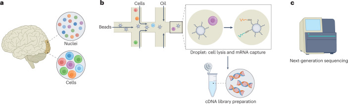
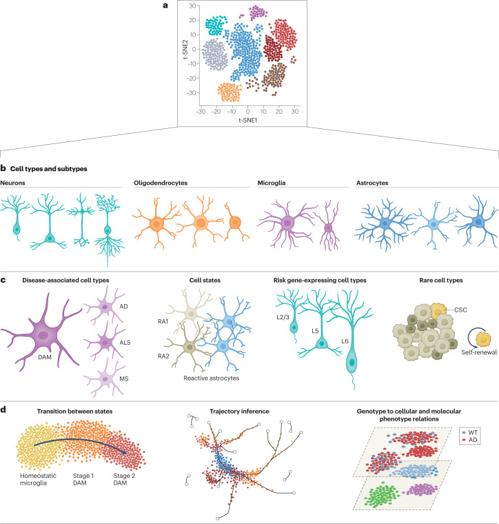
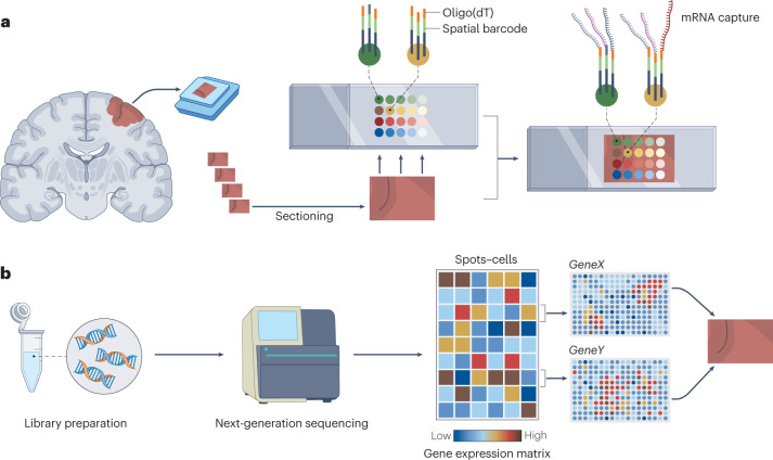
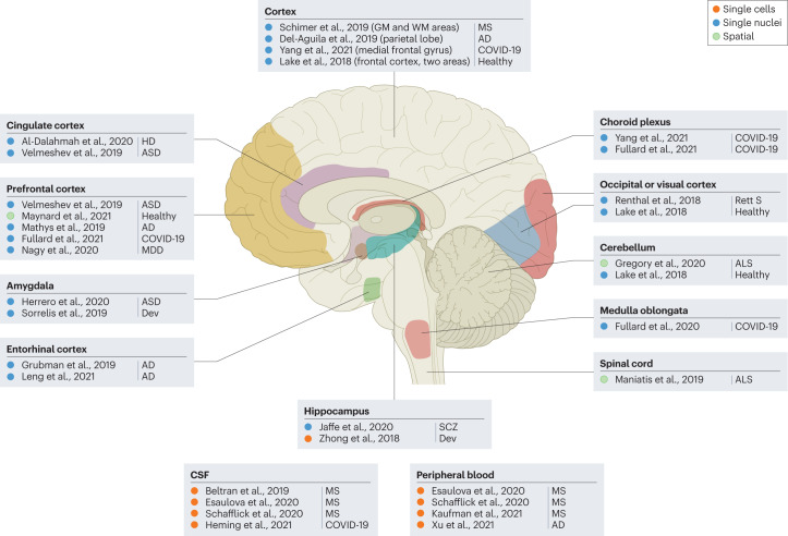
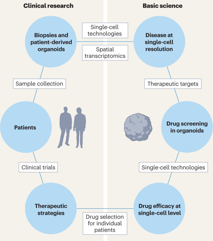
In the past decade, single-cell technologies have proliferated and improved from their technically challenging beginnings to become common laboratory methods capable of determining the expression of thousands of genes in thousands of cells simultaneously. The field has progressed by taking the CNS as a primary research subject - the cellular complexity and multiplicity of neuronal cell types provide fertile ground for the increasing power of single-cell methods. Current single-cell RNA sequencing methods can quantify gene expression with sufficient accuracy to finely resolve even subtle differences between cell types and states, thus providing a great tool for studying the molecular and cellular repertoire of the CNS and its disorders. However, single-cell RNA sequencing requires the dissociation of tissue samples, which means that the interrelationships between cells are lost. Spatial transcriptomic methods bypass tissue dissociation and retain this spatial information, thereby allowing gene expression to be assessed across thousands of cells within the context of tissue structural organization. Here, we discuss how single-cell and spatially resolved transcriptomics have been contributing to unravelling the pathomechanisms underlying brain disorders. We focus on three areas where we feel these new technologies have provided particularly useful insights: selective neuronal vulnerability, neuroimmune dysfunction and cell-type-specific treatment response. We also discuss the limitations and future directions of single-cell and spatial RNA sequencing technologies.


Conflict of interest statement

The authors declare no competing interests.

Figures

Fig. 1
Fig. 1. Experimental pipeline of a droplet-based single-cell RNA sequencing in a nutshell.
a, Generation of single-cell or single-nuclei suspensions from a tissue. b, Use of microfluidic device to encapsulate the individual cells or nuclei in nanolitre droplets with barcoded beads. Next steps include: cell lysis, capture of polyadenylated RNA, reverse transcription combined with the introduction of unique molecular identifiers and cell barcodes, and amplification and fragmentation of cDNA. c, Next-generation sequencing of the obtained cDNA library is performed on a standard platform, typically by solid-phase amplification.
Fig. 2
Fig. 2. Analysis of single-cell RNA sequencing data set can provide multiple types of information on cell types, states and their activation and enables inference of dynamic cellular processes.
a, High-dimensional single-cell RNA sequencing data can be visualized by using dimensionality reduction algorithms to reveal cell clusters. b, Cell clusters are assigned to specific cell types and subtypes on the basis of marker genes. c, Deeper analysis of single-cell transcriptomes can provide information about cell states and activation or uncover rare or new cell subtypes. d, Advanced algorithms and computational tools are designed to infer differentiation trajectories and transition states or compare phenotypes at the cellular level. AD, Alzheimer disease; ALS, amyotrophic lateral sclerosis; CSC, cancer stem cell; DAM, disease-associated microglia; MS, multiple sclerosis; RA, reactive astrocyte; t-SNE, t-distributed stochastic neighbour embedding; WT, wild type. Part d reprinted with permission from ref. , Elsevier.
Fig. 3
Fig. 3. Spatial transcriptomics: the principle and a workflow.
a, The tissue is subjected to cryosectioning on an mRNA capture slide, fixation and permeabilization to release RNA. The poly-A tail of the mRNA binds to an oligo(dT) ending fragment (single-stranded sequence of deoxythymines) on the capture DNA probes, which also contain embedded positional barcodes. b, After library preparation and sequencing, the computational analysis includes retrieval of the positional barcodes and tissue coordinates to reconstruct the relationship between transcripts and their locations.
Fig. 4
Fig. 4. Single-cell RNA-sequencing, single-nuclei RNA-sequencing and spatial transcriptomics studies reveal cellular and molecular heterogeneity in human neurological disorders.
Samples collected from the brain, spinal cord, cerebrospinal fluid (CSF) and peripheral blood have been used to analyse transcriptomes of thousands of cells and/or nuclei from individuals with multiple sclerosis (MS),–, Rett syndrome (Rett S), Alzheimer disease (AD),,,,, Huntington disease (HD), COVID-19 (refs. ), amyotrophic lateral sclerosis (ALS),, major depressive disorder (MDD), schizophrenia (SCZ) and autism spectrum disorders (ASD), and to compare them with healthy controls. Such an approach has also been used to compare different neurodevelopmental (Dev) stages,. GM, grey matter; WM, white matter.
Fig. 5
Fig. 5. Future directions for the application of single-cell and spatial transcriptomics in clinical use.
In the future, we expect that analysis of single cells from patients or patient-derived in vitro models will help to explore molecular mechanisms of diseases and define the spatial localization of rare cell types and cellular subpopulations emerging during disease. Furthermore, single-cell technologies will contribute to the discovery of new therapeutic targets. The efficacy of newly discovered drugs will then be tested in patient-derived in vitro models and monitored using single-cell technologies to define the cell‐type-specific responses of the patient to treatment, which can then be used to specify the best therapeutic strategy for the individual patient.

References

    1. Crino PB, Trojanowski JQ, Dichter MA, Eberwine J. Embryonic neuronal markers in tuberous sclerosis: single-cell molecular pathology. Proc. Natl Acad. Sci. USA. 1996;93:14152–14157. doi: 10.1073/pnas.93.24.14152. - DOI - PMC - PubMed
    1. Zeisel A, et al. Cell types in the mouse cortex and hippocampus revealed by single-cell RNA-seq. Science. 2015;347:1138–1142. doi: 10.1126/science.aaa1934. - DOI - PubMed
    1. Tasic B, et al. Adult mouse cortical cell taxonomy revealed by single cell transcriptomics. Nat. Neurosci. 2016;19:335–346. doi: 10.1038/nn.4216. - DOI - PMC - PubMed
    1. Eze UC, Bhaduri A, Haeussler M, Nowakowski TJ, Kriegstein AR. Single-cell atlas of early human brain development highlights heterogeneity of human neuroepithelial cells and early radial glia. Nat. Neurosci. 2021;24:584–594. doi: 10.1038/s41593-020-00794-1. - DOI - PMC - PubMed
    1. Tirosh I, et al. Single-cell RNA-seq supports a developmental hierarchy in human oligodendroglioma. Nature. 2016;539:309–313. doi: 10.1038/nature20123. - DOI - PMC - PubMed

Publication types