. 2023 Jul 11;148(2):109-123.
doi: 10.1161/CIRCULATIONAHA.122.062166. Epub 2023 May 18.

Retained Metabolic Flexibility of the Failing Human Heart

Affiliations

Retained Metabolic Flexibility of the Failing Human Heart

William D Watson et al. Circulation. .

Abstract

Background: The failing heart is traditionally described as metabolically inflexible and oxygen starved, causing energetic deficit and contractile dysfunction. Current metabolic modulator therapies aim to increase glucose oxidation to increase oxygen efficiency of adenosine triphosphate production, with mixed results.

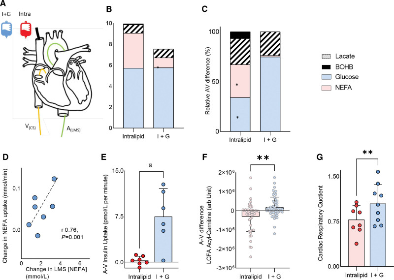
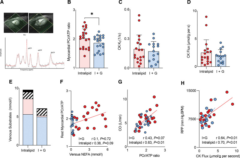
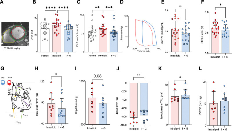
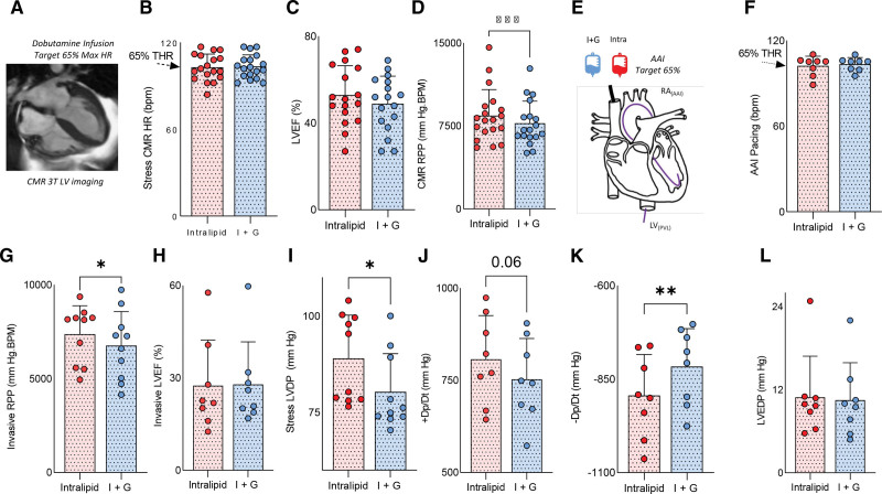
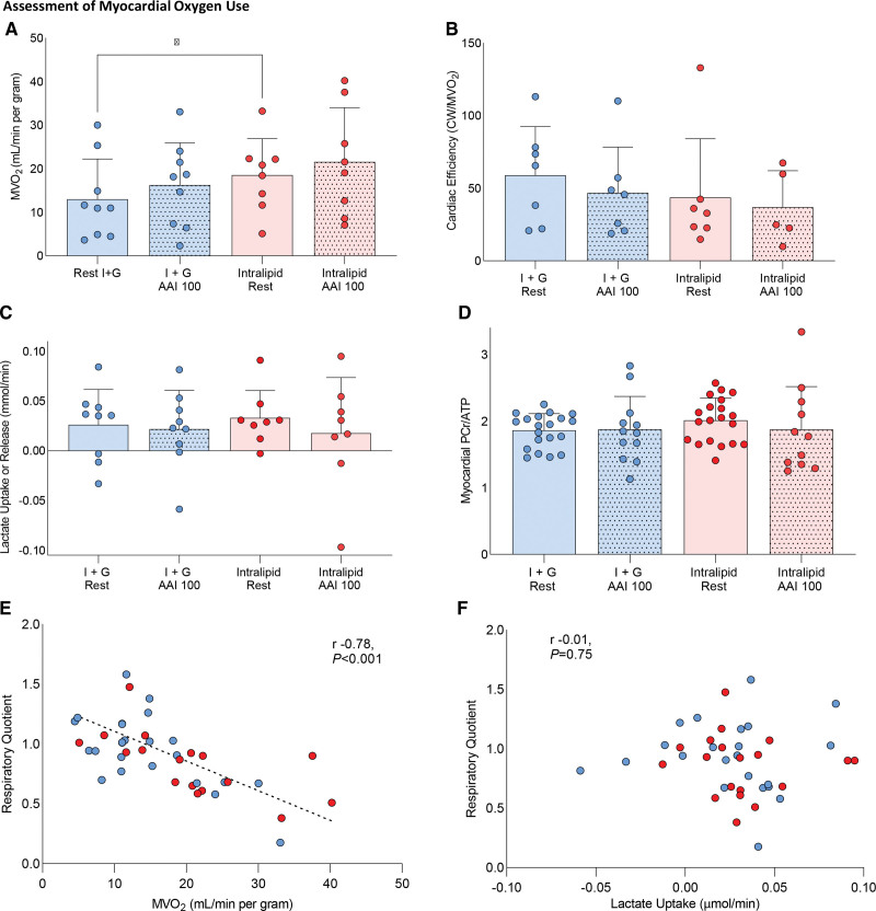
Methods: To investigate metabolic flexibility and oxygen delivery in the failing heart, 20 patients with nonischemic heart failure with reduced ejection fraction (left ventricular ejection fraction 34.9±9.1) underwent separate infusions of insulin+glucose infusion (I+G) or Intralipid infusion. We used cardiovascular magnetic resonance to assess cardiac function and measured energetics using phosphorus-31 magnetic resonance spectroscopy. To investigate the effects of these infusions on cardiac substrate use, function, and myocardial oxygen uptake (MVo2), invasive arteriovenous sampling and pressure-volume loops were performed (n=9).

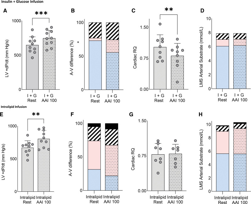
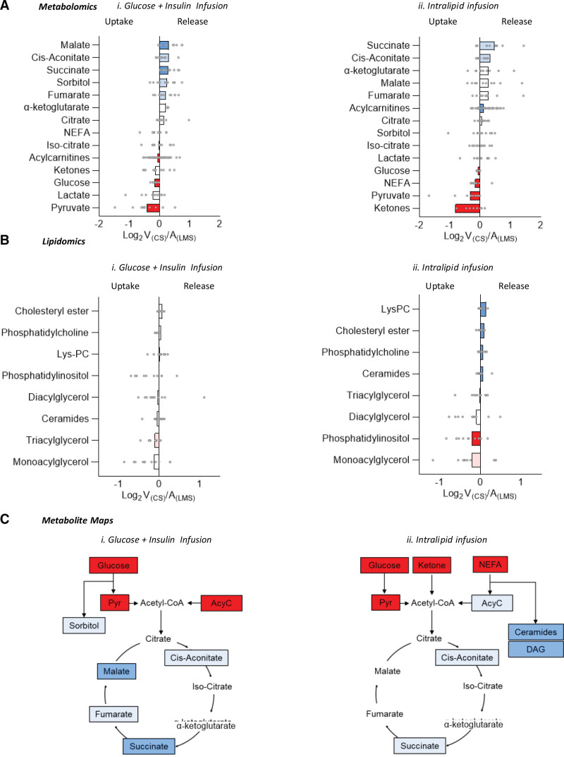
Results: At rest, we found that the heart had considerable metabolic flexibility. During I+G, cardiac glucose uptake and oxidation were predominant (70±14% total energy substrate for adenosine triphosphate production versus 17±16% for Intralipid; P=0.002); however, no change in cardiac function was seen relative to basal conditions. In contrast, during Intralipid infusion, cardiac long-chain fatty acid (LCFA) delivery, uptake, LCFA acylcarnitine production, and fatty acid oxidation were all increased (LCFA 73±17% of total substrate versus 19±26% total during I+G; P=0.009). Myocardial energetics were better with Intralipid compared with I+G (phosphocreatine/adenosine triphosphate 1.86±0.25 versus 2.01±0.33; P=0.02), and systolic and diastolic function were improved (LVEF 34.9±9.1 baseline, 33.7±8.2 I+G, 39.9±9.3 Intralipid; P<0.001). During increased cardiac workload, LCFA uptake and oxidation were again increased during both infusions. There was no evidence of systolic dysfunction or lactate efflux at 65% maximal heart rate, suggesting that a metabolic switch to fat did not cause clinically meaningful ischemic metabolism.

Conclusions: Our findings show that even in nonischemic heart failure with reduced ejection fraction with severely impaired systolic function, significant cardiac metabolic flexibility is retained, including the ability to alter substrate use to match both arterial supply and changes in workload. Increasing LCFA uptake and oxidation is associated with improved myocardial energetics and contractility. Together, these findings challenge aspects of the rationale underlying existing metabolic therapies for heart failure and suggest that strategies promoting fatty acid oxidation may form the basis for future therapies.

Keywords: adenosine triphosphate; heart failure; magnetic resonance spectroscopy; metabolism.


Conflict of interest statement

Disclosures None.

Figures

Figure 1.
Figure 1.
Assessment of cardiac substrate flexibility during altered substrate supply. A, Schematic of the experimental setup. B, Left main stem (LMS) arterial substrate concentrations. C, Relative arteriovenous (AV) difference of nonesterified free fatty acids (NEFA), glucose, lactate, and β-hydroxybutyrate (BOHB). D, Correlation between LMS NEFA concentration and uptake. E, Cardiac insulin uptake. F, Long-chain fatty acid (LCFA) acylcarnitines. G, Cardiac respiratory quotient (*P<0.05; **P<0.01; ***P<0.001). Data are presented as mean with SD error bars unless stacked. A(LMS) indicates arterial left main stem sample; and V(CS), venous coronary sample.
Figure 2.
Figure 2.
Assessment of cardiac substrate flexibility during increased workload with insulin+glucose infusion and Intralipid infusion. Assessment of cardiac substrate flexibility during increased workload (atria–atria–interrupt pacing 100 beats per minute [AAI 100]) during insulin+glucose (I+G) infusion (A through D) and Intralipid infusion (E through H) detailing relative arteriovenous difference of nonesterified free fatty acids, glucose, lactate, β-hydroxybutyrate (BOHB), left ventricle (LV)+change in pressure/change in time (dp/dt), and cardiac respiratory quotient (RQ). Spotted fill indicates stress measurement (*P<0.05; **P<0.01; ***P<0.001). Data are presented as mean with SD error bars unless stacked. In stacked charts, blue represents lipid; red, glucose; stripes, lactate; and black, ketone. LMS indicates left main stem.
Figure 3.
Figure 3.
Assessment of myocardial energetics at rest. A, 31P magnetic resonance spectroscopy. B, Phosphocreatine/adenosine triphosphate ratio (PCr/ATP). C, Creatine kinase (CK) first-order rate constant (CK kf). D, CK flux. E, Venous circulating substrates. F, The relationship between PCr/ATP and venous fatty acid (nonesterified free fatty acids [NEFA]) and cardiac output (CO; G). H, The relationships between CK flux and rate pressure product (RPP). Spotted indicates stress measurement (*P<0.05). Data are presented as mean with SD error bars. I+G indicates insulin+glucose.
Figure 4.
Figure 4.
Substrate manipulation and ventricular function at rest. A, Contouring. B, Left ventricular ejection fraction (LVEF). C, Stroke volume. D, An example pressure–volume loop. E, End systolic pressure–volume relationship (ESPVR). F, Stroke work. G, A graphic of the invasive experiment. H, Left ventricular developed pressure (LVDP). I, change in pressure/change in time (+dp/dt). J, -Dp/Dt. K, Isovolumetric tau. L, Left ventricular end diastolic pressure (LVEDP; *P<0.05; **P<0.01; ***P<0.001; ****P<0.0001). Data are presented as mean with SD error bars. Throughout, Intralipid data in red; insulin+glucose (I+G) data, in blue. A(LMS) indicates arterial left main stem sample; CMR, cardiac magnetic resonance; LV(PVL), left ventricle (pressure volume loop); and V(CS), venous coronary sample.
Figure 5.
Figure 5.
Substrate manipulation and ventricular function during stress. A, An example cardiac magnetic resonance (CMR) image. B, Dobutamine heart rate (HR), 65% target HR (THR) is shown. C, Left ventricular (LV) ejection fraction (LVEF). D, Rate pressure product (RPP). E, A graphic of the invasive stress experiment. F, HR during atria–atria–interrupt (AAI) pacing. G, RPP during AAI. H, LVEF by pressure–volume loop. I, Stress LVDP. J, Change in pressure/change in time (dp/dt). K, −dp/dt. L, LV end diastolic pressure (LVEDP; *P<0.05; **P<0.01; ***P<0.001; ****P<0.0001). Data are presented as mean with SD error bars. LV(PVL) indicates left ventricle (pressure volume loop); and RA, right atrium.
Figure 6.
Figure 6.
Assessment of the effects of substrate manipulation and variable workload on myocardial oxygen use. A, Myocardial oxygen use (MVo2 mL/min per gram). B, Cardiac efficiency (cardiac work/MVo2) changes. C, Cardiac lactate uptake. D, Myocardial phosphocreatine/adenosine triphosphate ratio (PCr/ATP) shown for the groups, along with the relationship between respiratory quotient and MVo2 (E) and myocardial lactate uptake (F). Dotted fill pattern demotes acquired during stress (*P<0.05). All analyses are repeated-measures ANOVA with Bonferroni correction. Correlation statistics are for Pearson R. Dotted fill denotes stress (atria–atria–interrupt [AAI] or dobutamine); plain denotes rest. Data presented as mean with SD error bars.
Figure 7.
Figure 7.
Arteriovenous metabolomics of cardiac uptake and release of substrates and tricarboxylic acid cycle intermediates, arteriovenous lipidomic assessment, and metabolic maps demonstrating the relative position in the tricarboxylic acid cycle of influxed and effluxed metabolites during insulin+glucose infusion and Intralipid infusion. A, Arteriovenous metabolomics of cardiac uptake/release of substrates and tricarboxylic acid cycle intermediates during (i) insulin+glucose (I+G) infusion and (ii) Intralipid infusion. B, Arteriovenous lipidomic assessment during (i) I+G infusion and (ii) Intralipid infusion. C, Metabolic maps demonstrating the relative position in the tricarboxylic acid cycle of influxed/effluxed metabolites during (i) I+G infusion and (ii) Intralipid infusion. Red denotes cardiac uptake (dark red, P<0.05; light red, P<0.1); blue, cardiac release (dark blue, P<0.05; light blue, P<0.1). All P values are adjusted for false discovery rate with Benjamini-Hochberg correction. A(LMS) indicates arterial left main stem sample; LysPC, lysophosphatidylcholine; NEFA, nonesterified free fatty acid; and V(CS), venous coronary sample.

References

    1. Peterzan MA, Lygate CA, Neubauer S, Rider OJ. Metabolic remodeling in hypertrophied and failing myocardium: a review. Am J Physiol Heart Circ Physiol. 2017;313:H597–H616. doi: 10.1152/ajpheart.00731.2016 - PubMed
    1. Doenst T, Nguyen TD, Abel ED. Cardiac metabolism in heart failure: implications beyond ATP production. Circ Res. 2013;113:709–724. doi: 10.1161/CIRCRESAHA.113.300376 - PMC - PubMed
    1. Heather LC, Cole MA, Lygate CA, Evans RD, Stuckey DJ, Murray AJ, Neubauer S, Clarke K. Fatty acid transporter levels and palmitate oxidation rate correlate with ejection fraction in the infarcted rat heart. Cardiovasc Res. 2006;72:430–437. doi: 10.1016/j.cardiores.2006.08.020 - PubMed
    1. Osorio JC, Stanley WC, Linke A, Castellari M, Diep QN, Panchal AR, Hintze TH, Lopaschuk GD, Recchia FA. Impaired myocardial fatty acid oxidation and reduced protein expression of retinoid X receptor-alpha in pacing-induced heart failure. Circulation. 2002;106:606–612. doi: 10.1161/01.cir.0000023531.22727.c1 - PubMed
    1. Davila-Roman VG, Vedala G, Herrero P, de las Fuentes L, Rogers JG, Kelly DP, Gropler RJ. Altered myocardial fatty acid and glucose metabolism in idiopathic dilated cardiomyopathy. J Am Coll Cardiol. 2002;40:271–277. doi: 10.1016/s0735-1097(02)01967-8 - PubMed

Publication types