. 2023 Jul 4;148(1):47-67.
doi: 10.1161/CIRCULATIONAHA.123.063760. Epub 2023 May 18.

INKILN is a Novel Long Noncoding RNA Promoting Vascular Smooth Muscle Inflammation via Scaffolding MKL1 and USP10

Affiliations

INKILN is a Novel Long Noncoding RNA Promoting Vascular Smooth Muscle Inflammation via Scaffolding MKL1 and USP10

Wei Zhang et al. Circulation. .

Abstract

Background: Activation of vascular smooth muscle cell (VSMC) inflammation is vital to initiate vascular disease. The role of human-specific long noncoding RNAs in VSMC inflammation is poorly understood.

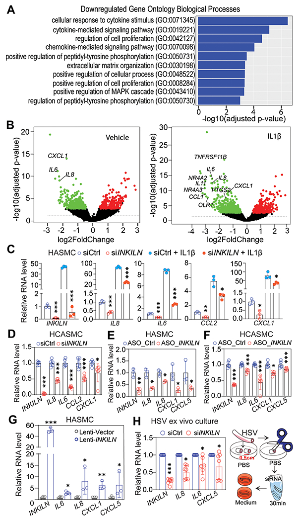
Methods: Bulk RNA sequencing in differentiated human VSMCs revealed a novel human-specific long noncoding RNA called inflammatory MKL1 (megakaryoblastic leukemia 1) interacting long noncoding RNA (INKILN). INKILN expression was assessed in multiple in vitro and ex vivo models of VSMC phenotypic modulation as well as human atherosclerosis and abdominal aortic aneurysm. The transcriptional regulation of INKILN was verified through luciferase reporter and chromatin immunoprecipitation assays. Loss-of-function and gain-of-function studies and multiple RNA-protein and protein-protein interaction assays were used to uncover a mechanistic role of INKILN in the VSMC proinflammatory gene program. Bacterial artificial chromosome transgenic mice were used to study INKILN expression and function in ligation injury-induced neointimal formation.

Results: INKILN expression is downregulated in contractile VSMCs and induced in human atherosclerosis and abdominal aortic aneurysm. INKILN is transcriptionally activated by the p65 pathway, partially through a predicted NF-κB (nuclear factor kappa B) site within its proximal promoter. INKILN activates proinflammatory gene expression in cultured human VSMCs and ex vivo cultured vessels. INKILN physically interacts with and stabilizes MKL1, a key activator of VSMC inflammation through the p65/NF-κB pathway. INKILN depletion blocks interleukin-1β-induced nuclear localization of both p65 and MKL1. Knockdown of INKILN abolishes the physical interaction between p65 and MKL1 and the luciferase activity of an NF-κB reporter. Furthermore, INKILN knockdown enhances MKL1 ubiquitination through reduced physical interaction with the deubiquitinating enzyme USP10 (ubiquitin-specific peptidase 10). INKILN is induced in injured carotid arteries and exacerbates ligation injury-induced neointimal formation in bacterial artificial chromosome transgenic mice.

Conclusions: These findings elucidate an important pathway of VSMC inflammation involving an INKILN/MKL1/USP10 regulatory axis. Human bacterial artificial chromosome transgenic mice offer a novel and physiologically relevant approach for investigating human-specific long noncoding RNAs under vascular disease conditions.

Keywords: RNA, long noncoding; inflammation; mice, transgenic; myocytes, smooth muscle; ubiquitination.


Conflict of interest statement

Disclosures None.

Figures

Figure 1.
Figure 1.. INKILN expression correlates with VSMC phenotypic modulation and vascular disease.
A. RNA-seq analysis revealed numerous coding (Gray dots) and noncoding genes (red dots) regulated by MYOCD in human coronary artery smooth muscle cells (HCASMCs) (n=2). B-D. qRT-PCR validation of the downregulation of INKILN in HCASMCs transduced with Ad-MYOCD relative to Ad-empty (B, n=3), differentiated HCASMCs induced by either TGFβ (2 ng/ml) (C, n=3) or conditioned SMC differentiation medium (SMD) versus growth medium (SMG) (D, n=3). E. qRT-PCR (left) and semi-qRT-PCR (right) analysis of the indicated genes in uncultured versus 2 weeks ex vivo cultured human saphenous vein (HSV) segments from the same patients (n=7 patients). F. qRT-PCR (left) and semi-qRT-PCR (right) analysis of the indicated genes in uncultured HSV versus primary cultured SMCs dispersed from fresh HSV tissues (HSVSMCs) (n=6). G. UCSC genome browser screenshot of the INKILN gene locus with combined single nucleus (sn) ATAC-seq libraries from healthy versus diseased coronary artery (CA) human samples (n=41). Healthy CA: patient has no evidence of atherosclerosis and samples are lesion-free; Athero I CA: patient has evidence of atherosclerosis, but samples are lesion-free; Atherosclerosis II CA: patient has evidence of atherosclerosis and sample contains lesion. H and I. qRT-PCR assessment of the indicated genes in human atherosclerotic plaque (Athero) versus non-plaque (Non-athero) regions from the same patients (H, n=8 patients), and abdominal aortic aneurysm (AAA) tissues (n=24 patients) relative to healthy control aortas (Control) from organ donors (H, n=6 donors). J. Representative images of the overview for the colorimetric ACTA2 (brown) immunohistochemistry staining of human AAA tissues and Immuno-RNA FISH for INKILN (Red) and a VSMC marker ACTA2 (Green) in the rectangle marked neointimal region (see overview) of human AAA vessels (n=5 patients). Arrows indicate specific INKILN signal. B-D, and F, unpaired t-test; E, paired t-test; H and I, Mann-Whitney test. *p<0.05, ** p <0.01, *** p <0.0001, ns, not significant.
Figure 2.
Figure 2.. INKILN is induced by proinflammatory stimuli through the NF-κB/p65-dependent pathway.
A. INKILN and its neighboring gene CXCL8 (IL8) expression in primary HSVSMCs ± IL1α and PDGF mined from the RNA-seq dataset we published. B. qRT-PCR analysis of INKILN expression in HSVSMCs induced with IL1α (10 ng/ml) and PDGF (20 ng/ml) relative to vehicle control (n=3). C-F. qRT-PCR assay for the indicated genes in HCASMCs ±TNFα (10 ng/ml) for 48 hours (C), HCASMCs ± IL1β (4 ng/ml) (D), human aortic SMCs (HASMCs) ± TNFα (10 ng/ml) (E) or IL1β (4 ng/ml) (F) for 24 hours (n=3). G. qRT-PCR for INKILN expression in HASMCs stimulated by IL1β (4 ng/ml) for the indicated time points (n=3). H. HASMCs were induced with IL1β (4 ng/ml) for 24 hours followed by treatment with BAY11-7082 (10 μM) for 24 hours before RNA extraction for qRT-PCR of the indicated genes (n=6). I. qRT-PCR analysis of INKILN in HASMCs transduced with Ad-IKKβ or vector control adenovirus (Ad-Empty) with the same dose (MOI=30) for 72 hours (n=3). J. Chromatin Immunoprecipitation (ChIP)-qPCR validation of p65 binding to the predicted NF-κB site within the proximal INKILN promoter in HASMs induced by IL1β or vehicle control for 15 minutes (n=3). K. Schematic of luciferase reporter of the putative −1.4 kb INKILN proximal promoter containing a predicted NF-κB site and the truncated −1.18 kb reporter lacking this site, and luciferase assays for the −1.4 kb promoter of INKILN and the truncated reporter in HEK293 cells induced by TNFα (10 ng/ml) for 6 hours (n=3). B, one-way ANOVA followed by a Bonferroni test; C-F, I, K (left), unpaired t-test; H, Brown-Forsythe and Welch ANOVA test followed by Dunnett’s multiple comparison test; J, and K(right), two-way ANOVA followed by a Tukey’s post hoc test. * p <0.05, ** p<0.01, *** p<0.0001, ns, not significant.
Figure 3.
Figure 3.. INKILN positively regulates proinflammatory gene expression.
A. The top 10 enriched Gene Ontology (GO) biological process terms downregulated by siINKILN in HASMCs under the IL1β-induced condition are shown (false discovery rate (FDR) adjusted p<0.05 and absolute log2FoldChange). Individual GO terms were sorted by adjusted p values. B. Volcano plot depicts the differentially expressed genes in HASMCs ± IL1β treated with siINKILN versus siCtrl (sicontrol). C-F. qRT-PCR validation of the reduced expression of the indicated pro-inflammatory genes upon INKILN depletion in HASMCs ± IL1β (C, n=3) and growing HCASMCs (D, n=6) using siINKILN versus siCtrl (n=3) or FANA Antisense Oligonucleotides (ASO) to INKILN (ASO_INKILN) versus ASO control (ASO_Ctrl) in growing HASMCs (E, n=3) and HCASMCs (F, n=3). G. Growing HASMCs transduced with the same amount of lentivirus carrying the INKILN (Lenti-INKILN) or lentivirus negative control (Lenti-vector) for 72 hours before RNA extraction for qRT-PCR of the indicated proinflammatory genes (n=3). H. HSV segments incubated with siINKILN or siCtrl for 30 minutes at the dose of 25 nM followed by ex vivo culture for 3 days before total RNA isolation for qRT-PCR of the indicated genes (each dot represents the average value from 3 separate segments from the same patient, n=6 patients). C-G, unpaired t-test; H, paired t-test. * p<0.05, ** p<0.01, *** p<0.0001, ns, not significant.
Figure 4.
Figure 4.. INKILN interacts with MKL1 in the cytoplasm of VSMCs.
A. Representative qRT-PCR analysis of INKILN and the indicated control genes in total RNA from the fractionated cytosolic and nuclear compartments in HCASMCs (n=3 independent experiments). B. RNA-FISH for INKILN (red) and PPIB (green) and DAPI (blue) staining in growing HCASMCs and the quantitation of the copy number of INKILN per cell (n=12 fields with 39 cells for siCtrl and n=17 fields with 71 cells for siINKILN from 3 biological replicates quantitated). C. In vitro RNA pulldown using biotinylated sense INKILN and antisense INKILN RNA showed an enriched band between 150kD and 250kD with sense INKILN by silver staining (red rectangle), which was validated as MKL1 protein by western blot (below). Representative images shown (n=3). D-E. Representative RNA Immunoprecipitation (RIP)-qPCR in HCASMC (D) and human rhabdomyosarcoma (RD) cells (E) showed an enrichment of INKILN from RNA precipitates by MKL1, but not p65 antibodies (n=3 independent experiments). F-G. Representative immuno-RNA-FISH for INKILN (red) and MKL1 protein (green) in HCASMCs (F, G) and the quantitation of the co-localization between INKILN and MKL1 protein by Pearson correlation coefficient analysis (G). PPIB mRNA was used as a negative control which fails to co-localize with MKL1 protein (quantitation was from 4 cells of 1 representative experiment out of 3 independent experiments). H. Representative immuno-RNA-FISH for INKILN and MKL1 protein in HCASMCs treated with Jasplakinolide (Jpk) for 6 hours or TGFβ for 24 hours to induce MKL1 nuclear translocation, and the quantitation of the co-localization of INKILN with MKL1 by Pearson correlation coefficient analysis under both stimulation conditions relative to their individual vehicle controls (quantitation was from 4 cells of 1 representative experiment out of 3 independent experiments). Scale Bar =20µm. B, G, and H, unpaired t-test; D and E, one-way ANOVA followed by a Dunnett’s test. * p<0.05, ** p<0.01, *** p<0.0001, ns, not significant.
Figure 5.
Figure 5.. Loss of INKILN suppresses MKL1/p65-mediated activation of the proinflammatory gene program.
A. qRT-PCR analysis of the expression levels of the indicated proinflammatory genes in HCASMCs treated with same amount of lentivirus carrying short hairpin RNA to MKL1 (Lenti-shMKL1) or siRNA SMART POOL to MKL1 (siMKL1) versus their individual controls (Lenti-shCtrl or siCtrl) (n=3). B-C. Representative western blot of the phosphorylated p65 (pp65) level in HCASMCs transduced with Lenti-shMKL1 versus Lenti-shCtrl (B, n=3) or Adenovirus carrying MKL1 transcript (Ad-MKL1) versus Ad-empty control (Ad-empty) for 48 hours before protein extraction for western blot of the indicated proteins (C, n=4) and the respective quantitation. D. Representative western blot of fractionated proteins from the indicated cellular compartments in HCASMCs depleted by siINKILN for 48 hours followed by IL1β stimulation for 24 hours and the quantitation (n=4). E. Representative immunofluorescence staining for p65 protein in HASMCs treated with siINKILN or siCtrl for 48 hours prior to IL1β induction for 24 hours and the quantitation (n=3). F. Immunofluorescence staining for MKL1 in HASMCs treated with siINKILN versus siCtrl for 48 hours followed by IL1β induction for 24 hours (n=3 with indicated total cell numbers). G. Luciferase assay for NF-κB reporter activity in HASMCs depleted by siINKILN for 48 hours followed by TNFα (10 ng/ml) simulation for 6 hours (n=3). A-C, unpaired t-test; D, Brown-Forsythe and Welch ANOVA test followed by Dunnett’s multiple comparison test; E and F, Mann Whitney test; G, two-way ANOVA followed by a Tukey’s post hoc test. * p<0.05, ** p<0.01, *** p<0.0001, ns, not significant.
Figure 6.
Figure 6.. Loss of INKILN reduces MKL1 protein stability via enhancing ubiquitination proteasome degradation.
A. HASMCs were transfected with siINKILN or siCtrl for 48 hours prior to protein extraction for p65 immunoprecipitation followed by western blotting analysis of the indicated proteins.1/100 amount of total cell lysates were used as input control. Representative western blot images for the indicated proteins (n=5). B-D. HCASMCs (B, n=4), HASMCs (C, n=4), and RD cells (D, n=3) were treated with siINKILN or siCtrl for 72 hours and protein lysates were used for western blot analysis of MKL1. Representative western blot images (B-D, top) and the quantitation (B-D, bottom). E. qRT-PCR of MKL1 mRNA expression after siRNA-mediated INKILN gene knockdown in HCASMCs (n=3). F. HASMCs treated with 5μM MG132 for the indicated time before protein extraction for western blot of MKL1. Representative western blot (top) and the quantitation at 20 hours after the treatment of MG132 (bottom) (n=5). G. HASMCs treated with siRNA for 48 hours followed by MG132 (5 μM) for 20 hours prior to protein extraction for western blot of MKL1. Representative western blot image (top) and the quantification (bottom) (n=4). H. HCASMCs treated with siINKILN versus siCtrl for 48 hours followed by MG132 (5 μM) treatment for 20 hours prior to protein extraction for immunoprecipitation of MKL1 and western blot of ubiquitin. Representative images shown (n=4). B and E, one-way ANOVA followed by a Bonferroni test; C, D, and F, unpaired t-test; G, two-way ANOVA followed by a Bonferroni test. * p<0.05, ** p<0.01, *** p<0.0001, ns, not significant.
Figure 7.
Figure 7.. INKILN facilitates the interaction between MKL1 and USP10.
A. Representative image of co-immunoprecipitation of MKL1 followed by western blot of the indicated proteins in HCASMCs (n=4). B. Representative co-immunofluorescence staining of MKL1 and USP10 in HCASMCs (n=3). Scale Bar=20μm. C,D. Representative western blot of the indicated proteins in HCASMCs treated with siRNA to USP10 (C), or adenovirus overexpressing USP10 (D) versus their individual controls and the quantitation of MKL1 protein levels (n=11). E. HCASMCs were treated with siRNA-INKILN for 72 hours and the protein levels of USP10 were detected by western blot (n=6). F. HCASMCs were treated with siRNA-INKILN for 72 hours prior to immunoprecipitation of MKL1 and western blot of the indicated proteins. Representative image and the quantitation of 8 biological replicates from 4 independent experiments. G, Representative image of proximity ligation assay (PLA) for MKL1 and USP10 in HCASMCs, and quantitation of PLA punctae shown (17 fields from n=5 independent experiments). H. qRT-PCR of the indicated genes from the RNA pools precipitated by USP10 antibody in HCASMCs (n=3). Unpaired t-test for all the comparisons. * p<0.05, ** p<0.01, *** p<0.0001, ns, not significant.
Figure 8.
Figure 8.. INKILN expression in BAC transgenic mice and its influence on neointimal formation.
A. CRISPR-LRS mapped a single integration locus for human INKILN. The integration locus, indicated by a grey box, spanned 32,808,215bp - 32,828,044bp on mouse chromosome 11 (mm10), disrupting testis-specific protein-coding gene, Smim23. INKILN BAC transgenes (brown rectangles), integrated in a tandem head to tail fashion accompanied with BAC cloning vector sequence (red boxes). B. qPCR determined ~2 transgene copies for human INKILN (+/tg, n=6) with data normalized to internal control locus. Itga8-CreERT2 mice (n=2) served as calibrator for one copy of a transgene.84 Values graphed as mean ± SEM. C. qRT-PCR of INKILN for the uncultured versus 3 days ex vivo cultured aorta segments from WT and INKILN transgenic (Tg) mice (n=6). D. qRT-PCR of INKILN for unligated versus 1 week ligated carotid arteries from WT and Tg mice (n=3). E. Representative RNA FISH image for INKILN transcripts in unligated versus 4 week ligated carotid arteries from WT and Tg mice (n=3). F, G. Representative whole mount of 4 week ligated carotid arteries from WT versus Tg mice (F, left), the H&E staining of sections at different levels (F, right), and the quantitation of neointimal formation (G, n=13 for WT and n=15 for Tg). Representative images of immunofluorescence staining (H) for the indicated proteins on cross sections of ligated carotid arteries from WT and Tg mice and the quantitation of the fluorescence positive area over the total nuclei at neointima and media (n= 5 mice, 1 section/mouse at level 3). I. Western blot of the indicated proteins in unligated and ligated carotid arteries from WT versus INKILN Tg mice and the quantitation (n=6). NI, Neointima; M, Media; UL, unligated carotids; L, ligated carotids. G and I, Mann-Whitney test; H, paired t-test. *p<0.05, **p<0.01, ns, not significant. J. Working model of INKILN activating VSMC inflammation. Inflammation induces INKILN expression, which inhibits MKL1 ubiquitin proteasome degradation via USP10 and enhances both MKL1 and p65 nuclear translocation, resulting in the increased nuclear interaction of MKL1 with p65 and subsequent transactivation of the proinflammatory gene program.

Update of

References

    1. Zanoli L, Briet M, Empana JP, Cunha PG, Mäki-Petäjä KM, Protogerou AD, Tedgui A, Touyz RM, Schiffrin EL, Spronck B, Bouchard P, Vlachopoulos C, Bruno RM and Boutouyrie P. Vascular consequences of inflammation: a position statement from the ESH Working Group on Vascular Structure and Function and the ARTERY Society. J Hypertens. 2020;38:1682–1698. - PMC - PubMed
    1. Shah PK. Inflammation, neointimal hyperplasia, and restenosis: as the leukocytes roll, the arteries thicken. Circulation. 2003;107:2175–7. - PubMed
    1. Williams JW, Huang LH and Randolph GJ. Cytokine Circuits in Cardiovascular Disease. Immunity. 2019;50:941–954. - PMC - PubMed
    1. Liberale L, Ministrini S, Carbone F, Camici GG and Montecucco F. Cytokines as therapeutic targets for cardio- and cerebrovascular diseases. Basic Res Cardiol. 2021;116:23. - PMC - PubMed
    1. Nguyen MT, Fernando S, Schwarz N, Tan JT, Bursill CA and Psaltis PJ. Inflammation as a Therapeutic Target in Atherosclerosis. J Clin Med. 2019;8. - PMC - PubMed

Publication types

LinkOut - more resources