Review
. 2023 Sep 28;67(6):919-928.
doi: 10.1042/EBC20220242.

Perivascular tumor-associated macrophages and their role in cancer progression

Affiliations
Review

Perivascular tumor-associated macrophages and their role in cancer progression

Meriem Bahri et al. Essays Biochem. .

Abstract

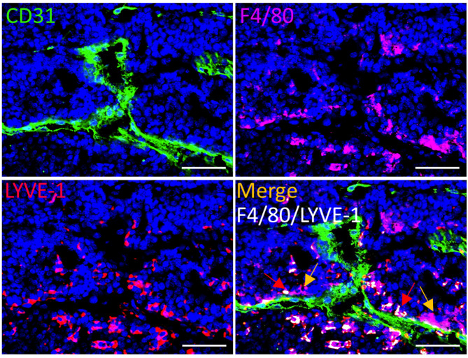
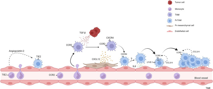
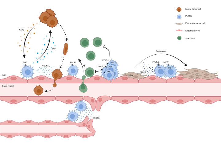
Perivascular (Pv) tumor-associated macrophages (TAMs) are a highly specialized stromal subset within the tumor microenvironment (TME) that are defined by their spatial proximity, within one cell thickness, to blood vasculature. PvTAMs have been demonstrated to support a variety of pro-tumoral functions including angiogenesis, metastasis, and modulating the immune and stromal landscape. Furthermore, PvTAMs can also limit the response of anti-cancer and anti-angiogenic therapies and support tumor recurrence post-treatment. However, their role may not exclusively be pro-tumoral as PvTAMs can also have immune-stimulatory capabilities. PvTAMs are derived from a monocyte progenitor that develop and localize to the Pv niche as part of a multistep process which relies on a series of signals from tumor, endothelial and Pv mesenchymal cell populations. These cellular communications and signals create a highly specialized TAM subset that can also form CCR5-dependent multicellular 'nest' structures in the Pv niche. This review considers our current understanding of the role of PvTAMs, their markers for identification, development, and function in cancer. The role of PvTAMs in supporting disease progression and modulating the outcome from anti-cancer therapies highlight these cells as a therapeutic target. However, their resistance to pan-TAM targeting therapies, such as those targeting the colony stimulating factor-1 (CSF1)-CSF1 receptor axis, prompts the need for more targeted therapeutic approaches to be considered for this subset. This review highlights potential therapeutic strategies to target and modulate PvTAM development and function in the TME.

Keywords: cancer; macrophages; metastasis; perivascular; polarization; tumor microenvironments.


Conflict of interest statement

The authors declare that there are no competing interests associated with the manuscript.

Figures

Figure 1
Figure 1. Models for TAM polarization
The dichotomy of the conventionally defined ‘M1’ and ‘M2’ macrophage polarization program, as pro- (M1) and anti- (M2) inflammatory stages of their phenotypic program, has served as a valuable framework for understanding and classifying the extremes of TAM phenotypes (left). However, a more inclusive ‘spectrum’ polarization model was proposed to enable the capturing of inter- and intra-tumoral TAM phenotypes identified (middle). Recent advances in our knowledge of TAM development, have highlighted that the most specialized TAM phenotypes develop in a multistep process and, as such, we propose a third ‘developmental’ model which encompasses both the heterogeneity of TAM phenotypes and the potential linkages of these phenotypes into developmental pathways within the TME (right). Although these models are most easily applied to monocyte-derived macrophages, it should be noted that self-renewing tissue-resident macrophages can also get incorporated into a growing tumor and contribute to TAM heterogeneity which are not reflected in the above schematics.
Figure 2
Figure 2. Evidence of TAM heterogeneity and nest formation in the Pv niche
Representative image of a frozen section of tumor from the spontaneous MMTV-PyMT murine model of breast cancer [66], stained with DAPI (nuclei;blue) and antibodies against F4/80 (magenta), LYVE-1 (red) and CD31 (green). Colocalizing pixels for F4/80 and LYVE-1 is shown in white. Red arrows highlight examples of LYVE-1+ PvTAMs and yellow arrows highlight examples of LYVE-1 PvTAMs within the TME. Scale bar: 25 µm.
Figure 3
Figure 3. Development of PvTAMs
Summary of the developmental pathways of PvTAMs highlighted in the manuscript text. TIE2+ PvTAMs can arise from TIE2 expressing monocytes recruited from the peripheral blood. TIE2+ PvTAMs are retained in the Pv niche through their interaction with angiopoietin-2 expressed by endothelial cells. Additionally, CCR2-expressing monocytes enter the tumor and develop into PvTAMs. Upon entering the tumor, TAMs respond to TGF-β derived from tumor cells which up-regulate CXCR4. CXCR4+ TAMs then migrate towards CXCL12 expressed by Pv mesenchymal cells to reach the Pv niche. IL-6 expressed by the endothelium polarizes PvTAMs to develop a LYVE-1+CCR5+HO-1+ phenotype. LYVE-1+ PvTAMs express CCL3 and CCL4 allowing the subset to communicate via CCR5 to form multicellular nests in the Pv niche. Black arrows denote differentiation and dashed arrows represent ligand/receptor interactions. It is currently unknown if LYVE-1+CCR5+HO-1+ PvTAM, CXCR4+, and TIE2+ PvTAM populations are related or if they are discrete phenotypic and functional populations.
Figure 4
Figure 4. PvTAM functions in the TME
Summary of the functions of PvTAMs highlighted in the manuscript text. PvTAMs can facilitate intravasation of Mena+ tumor cells into the circulation, where PvTAMs express EGF and tumor cells express CSF1 and participate in a cross-communication axis, which results in streaming of tumor cells to the Pv niche. PvTAMs facilitate intravasation of tumor cells through their secretion of VEGFA which results in temporary vessel leakiness. Additionally, PvTAMs are associated with an emerging role in modulating the immune landscape of the TME, where there are reports that populations of PvTAMs are associated with exclusion (Lyve1+HO1+ PvTAMs) and recruitment (FOLR2+ PvTAMs) of CD8+ T-cells into the tumor. LYVE-1+ PvTAMs orchestrate a selective expansion of a pericyte-like mesenchymal cell population through their expression of PDGF-C, creating a pro-angiogenic niche. PvTAMs, can facilitate neo-angiogenesis and anastomosis of blood vessels in the TME.

References

    1. Mantovani A., Marchesi F., Malesci A., Laghi L. and Allavena P. (2017) Tumour-associated macrophages as treatment targets in oncology. Nat. Rev. Clin. Oncol. 14, 399–416 10.1038/nrclinonc.2016.217 - DOI - PMC - PubMed
    1. DeNardo D.G., Barreto J.B., Andreu P., Vasquez L., Tawfik D., Kolhatkar N.et al. . (2009) CD4(+) T cells regulate pulmonary metastasis of mammary carcinomas by enhancing protumor properties of macrophages. Cancer Cell. 16, 91–102 10.1016/j.ccr.2009.06.018 - DOI - PMC - PubMed
    1. Muliaditan T., Caron J., Okesola M., Opzoomer J.W., Kosti P., Georgouli M.et al. . (2018) Macrophages are exploited from an innate wound healing response to facilitate cancer metastasis. Nat. Commun. 9, 2951 10.1038/s41467-018-05346-7 - DOI - PMC - PubMed
    1. Franklin R.A., Liao W., Sarkar A., Kim M.V., Bivona M.R., Liu K.et al. . (2014) The cellular and molecular origin of tumor-associated macrophages. Science 344, 921–925 10.1126/science.1252510 - DOI - PMC - PubMed
    1. Colegio O.R., Chu N.Q., Szabo A.L., Chu T., Rhebergen A.M., Jairam V.et al. . (2014) Functional polarization of tumour-associated macrophages by tumour-derived lactic acid. Nature 513, 559–563 10.1038/nature13490 - DOI - PMC - PubMed

Publication types