. 2023 Jun 28;89(6):e0059423.
doi: 10.1128/aem.00594-23. Epub 2023 May 18.

Environmental and Taxonomic Drivers of Bacterial Extracellular Vesicle Production in Marine Ecosystems

Affiliations

Environmental and Taxonomic Drivers of Bacterial Extracellular Vesicle Production in Marine Ecosystems

Steven J Biller et al. Appl Environ Microbiol. .

Abstract

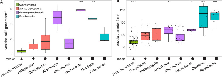
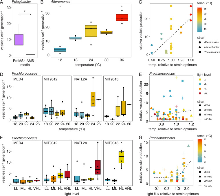
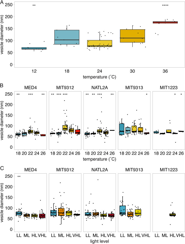
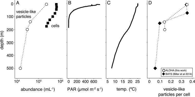
Extracellular vesicles are small (approximately 50 to 250 nm in diameter), membrane-bound structures that are released by cells into their surrounding environment. Heterogeneous populations of vesicles are abundant in the global oceans, and they likely play a number of ecological roles in these microbially dominated ecosystems. Here, we examine how vesicle production and size vary among different strains of cultivated marine microbes as well as explore the degree to which this is influenced by key environmental variables. We show that both vesicle production rates and vesicle sizes significantly differ among cultures of marine Proteobacteria, Cyanobacteria, and Bacteroidetes. Further, these properties vary within individual strains as a function of differences in environmental conditions, such as nutrients, temperature, and light irradiance. Thus, both community composition and the local abiotic environment are expected to modulate the production and standing stock of vesicles in the oceans. Examining samples from the oligotrophic North Pacific Gyre, we show depth-dependent changes in the abundance of vesicle-like particles in the upper water column in a manner that is broadly consistent with culture observations: the highest vesicle abundances are found near the surface, where the light irradiances and the temperatures are the greatest, and they then decrease with depth. This work represents the beginnings of a quantitative framework for describing extracellular vesicle dynamics in the oceans, which is essential as we begin to incorporate vesicles into our ecological and biogeochemical understanding of marine ecosystems. IMPORTANCE Bacteria release extracellular vesicles that contain a wide variety of cellular compounds, including lipids, proteins, nucleic acids, and small molecules, into their surrounding environment. These structures are found in diverse microbial habitats, including the oceans, where their distributions vary throughout the water column and likely affect their functional impacts within microbial ecosystems. Using a quantitative analysis of marine microbial cultures, we show that bacterial vesicle production in the oceans is shaped by a combination of biotic and abiotic factors. Different marine taxa release vesicles at rates that vary across an order of magnitude, and vesicle production changes dynamically as a function of environmental conditions. These findings represent a step forward in our understanding of bacterial extracellular vesicle production dynamics and provide a basis for the quantitative exploration of the factors that shape vesicle dynamics in natural ecosystems.

Keywords: Alcanivorax; Alteromonas; Dokdonia; Marinobacter; North Pacific Gyre; Pelagibacter; Polaribacter; Prochlorococcus; Thalassospira; extracellular vesicles.


Conflict of interest statement

The authors declare no conflict of interest.

Figures

FIG 1
FIG 1
Variation in extracellular vesicle properties across selected marine microbial genera. The plots indicate the measured differences in (A) vesicle production rates and (B) vesicle sizes. The Prochlorococcus data represent the median values from five strains that were grown at different light levels (Fig. 2). All cells were grown at 24°C, except Pelagibacter, which grew at 22°C. Shapes indicate the seawater-based medium that was required to support strain growth: Pro99 (triangle), ProMS* (diamond), ProMM (circle), or Pro99-PY (square). The colors represent the taxonomic groupings of microbes at the class level: Cyanophyceae (green), Alphaproteobacteria (red), Gammaproteobacteria (purple), or Flavobacteriia (blue). Asterisks indicate strains that differ significantly from the overall mean (Wilcoxon test; *, P ≤ 0.05; **, P ≤ 0.01; ****, P ≤ 0.0001).
FIG 2
FIG 2
Vesicle production rate variation with changes in environmental factors. (A) Pelagibacter vesicle production in two different media. (B) Vesicle release across growth temperatures in Alteromonas. (C) Relative changes in vesicle release rates across three different marine heterotrophs, relative to their strain-specific growth optima. (D) Vesicle production rates from four Prochlorococcus strains with changes in the growth temperature. (E) Relative vesicle production rates by Prochlorococcus strains, compared to the temperature that maximized the growth rate for each strain and light level (optimal = 1). (F) Vesicle production rates from four Prochlorococcus strains with changes in light irradiance. LL, low light; ML, medium light; HL, high light; VHL, very high light (Table S1). (G) Relative Prochlorococcus vesicle production rates as a function of the light level. Vesicle release is normalized, relative to the minimum measured value for each strain. The light levels are compared to the irradiance that resulted in the maximal growth rate for each strain (optimal = 1). The dashed line in panel C indicates the linear regression fit. The lines in panels E and G represent the change point regression fit. *, P < 0.05; Wilcoxon test for panels A and F; t test in panel B.
FIG 3
FIG 3
Vesicle size variation with changes in environmental factors. The measured vesicle diameters are shown for (A) Alteromonas macleodii MIT1002 across growth temperatures, (B) Prochlorococcus strains across growth temperatures, and (C) Prochlorococcus strains grown at different relative light irradiances. LL, low light; ML, medium light; HL, high light; VHL, very high light (Table S1). Asterisks indicate conditions that differ significantly from the overall strain mean diameter (Wilcoxon test; *, P ≤ 0.05; **, P ≤ 0.01; ***, P ≤ 0.001; ****, P ≤ 0.0001).
FIG 4
FIG 4
Distribution of vesicle-like particles in the North Pacific Subtropical Gyre. (A) Concentration of vesicle-like particles (open circles) and cells (bacteria and eukaryotes, filled squares) from Station ALOHA in June of 2014. Contextual environmental data are shown for (B) photosynthetically active radiation (PAR) levels and (C) temperature. (D) Relative ratio of vesicle-like particles to cells from Station ALOHA (this work, open circles) and the Bermuda-Atlantic Time Series Station (BATS, collected in December of 2012, filled diamond) (1).

References

    1. Biller SJ, Schubotz F, Roggensack SE, Thompson AW, Summons RE, Chisholm SW. 2014. Bacterial vesicles in marine ecosystems. Science 343:183–186. doi:10.1126/science.1243457. - DOI - PubMed
    1. Biller SJ, McDaniel LD, Breitbart M, Rogers E, Paul JH, Chisholm SW. 2017. Membrane vesicles in sea water: heterogeneous DNA content and implications for viral abundance estimates. ISME J 11:394–404. doi:10.1038/ismej.2016.134. - DOI - PMC - PubMed
    1. Biller SJ, Lundeen RA, Hmelo LR, Becker KW, Arellano AA, Dooley K, Heal KR, Carlson LT, Van Mooy BAS, Ingalls AE, Chisholm SW. 2022. Prochlorococcus extracellular vesicles: molecular composition and adsorption to diverse microbes. Environ Microbiol 24:420–435. doi:10.1111/1462-2920.15834. - DOI - PMC - PubMed
    1. Baeza N, Mercade E. 2021. Relationship between membrane vesicles, extracellular ATP and biofilm formation in Antarctic Gram-negative bacteria. Microb Ecol 81:645–656. doi:10.1007/s00248-020-01614-6. - DOI - PMC - PubMed
    1. Erdmann S, Tschitschko B, Zhong L, Raftery MJ, Cavicchioli R. 2017. A plasmid from an Antarctic haloarchaeon uses specialized membrane vesicles to disseminate and infect plasmid-free cells. Nat Microbiol 2:1446–1455. doi:10.1038/s41564-017-0009-2. - DOI - PubMed

Publication types

LinkOut - more resources