. 2023 May 18;75(1):41.
doi: 10.1186/s43044-023-00368-z.

A clinical case of successful palliative endovascular treatment of a patient with a single ventricle, mitral valve atresia, an intact atrial septum and persistent cardinal vein

Affiliations

A clinical case of successful palliative endovascular treatment of a patient with a single ventricle, mitral valve atresia, an intact atrial septum and persistent cardinal vein

Gorbatykh Artem et al. Egypt Heart J. .

Abstract

Background: Treatment of newborns with univentricular hemodynamics in combination with an anomaly of pulmonary venous return has the worst correction results in modern cardiac surgical papers. According to the data obtained by different authors, postoperative mortality in this cohort of patients varies from 41.7 to 53%. The presence of the venous outflow tract obstruction, as well as the serious condition of a newborn, is one of the main factors that increase the risk of death in the postoperative period.

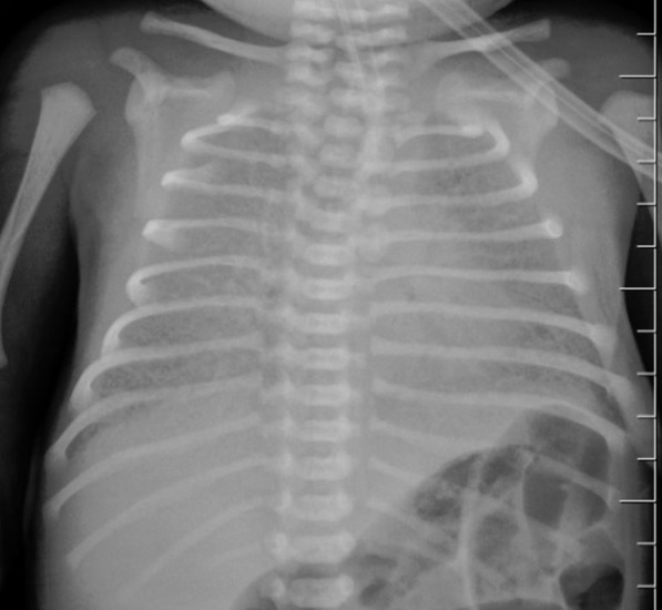
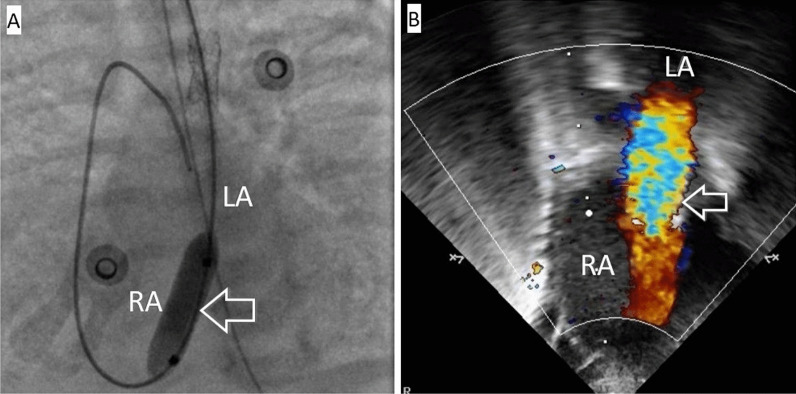
Case presentation: This article reveals a clinical case of a patient with a combined heart disease prenatally diagnosed in the form of a functionally single ventricle with a double outlet of the main vessels from it, mitral valve atresia, an intact atrial septum and an anomaly of venous return, when the blood outflow from the left atrium was carried out through a single fetal communication such as stenotic cardinal vein. In order to stabilize the patient's condition, the newborn urgently underwent stenting of the stenotic section of the cardinal vein. However, due to the lack of positive dynamics in the postoperative period, the child underwent repeated endovascular intervention and stenting of the intraoperatively created interatrial communication was performed. Taking into account the absence of obstruction of the outflow tract to the pulmonary artery, it was necessary to perform an open surgical intervention in a short time such as pulmonary artery banding.

Conclusions: Thus, palliative endovascular intervention in critically ill neonates with univentricular hemodynamics and anomalous pulmonary venous return can be considered as a method of choice that can become a new safer strategy for managing infants in order to stabilize the condition before the main stage of surgical intervention comes.

Keywords: Anomalous venous return; Endovascular palliative intervention.


Conflict of interest statement

The authors declare that they have no competing interests.

Figures

Fig. 1
Fig. 1
Chest X-ray, front view. Total infiltration of both lungs is determined mainly due to the compaction of the pulmonary interstitium
Fig. 2
Fig. 2
CT of chest. A Intact atrial septum (indicated by arrow). B Patent ductus arteriosus (indicated by arrow). C Cardinal vein (the only way outflow from the left atrium). D The aorta and pulmonary artery go from a single ventricle. Ao aorta, CV cardinal vein, IAS intact atrial septum, LA left atrium, MPA main pulmonary artery, PDA patent ductus arteriosus, RA right atrium, SV single ventricle
Fig. 3
Fig. 3
Anatomical representation of venous return anomaly at the primary form. A Schematic designation of venous outflow from the LA through a stenotic CV. B Angiography with stenosis zone visualization (indicated by the arrow) of the CV, anteroposterior view. Ao aorta, CV cardinal vein, IV innominate vein, IVC inferior vena cava, LA left atrium, MPA main pulmonary artery, RA right atrium, SV single ventricle, SVC superior vena cava
Fig. 4
Fig. 4
Anatomical representation of venous return anomaly after cardinal vein stenting. A Schematic representation of venous outflow from the left atrium after cardinal vein stenting. B The stent is implanted in the narrowed area (indicated by the arrow), anteroposterior view. C The area of ​​stenting (indicated by the arrow) is passable, the contrast agent freely enters the innominate vein, anteroposterior view. CV cardinal vein, IV innominate vein, IVC inferior vena cava, LA left atrium, RA right atrium, SV single ventricle; SVC, superior vena cava
Fig. 5
Fig. 5
Balloon dilatation of the AS under simultaneous X-ray and Echocardiography. A Balloon septostomy under angiographic control, the balloon (indicated by the arrow) is inflated in the area of ​​the atrial septal defect, anteroposterior view. B Echocardiography after balloon septostomy. Interatrial communication is 3 mm in size (indicated by an arrow). LA left atrium, RA right atrium
Fig. 6
Fig. 6
Anatomical representation of venous return abnormality after stenting of the CV and interatrial communication. A Schematic illustration of venous outflow from the LA after stenting of the CV and interatrial communication. B Stent is in the area of ​​the AS (indicated by an arrow), anteroposterior view. C The area of ​​stenting is passable, the contrast from the LA flows into the RA through the stented CV and the stented interatrial communication (indicated by the arrow), anteroposterior view. CV cardinal vein, IV innominate vein, IVC inferior vena cava, LA left atrium, RA right atrium, SV single ventricle, SVC superior vena cava

References

    1. Vanderlaan RD, Caldarone ChA. Surgical approaches to total anomalous pulmonary venous connection. Semin Thorac Cardiovasc Surg Pediatr Card Surg Annu. 2018;21:83–91. doi: 10.1053/j.pcsu.2017.11.010. - DOI - PubMed
    1. Kao CC, Hsieh CC, Cheng PJ, Chiang CH, Huang SY. Total anomalous pulmonary venous connection: from embryology to a prenatal ultrasound diagnostic update. J Med Ultrasound. 2017;25(3):130–137. doi: 10.1016/j.jmu.2017.08.002. - DOI - PMC - PubMed
    1. Kobayashi D, Forbes TJ, Aggarwal S. Palliative stent placement in vertical vein in a 1.4 kg infant with obstructed supracardiac total anomalous pulmonary venous connection. Catheter Cardiovasc. Interv. 2013;82(4):574–580. doi: 10.1002/ccd.24632. - DOI - PubMed
    1. George RS, Lozier JS, Bocks ML. Palliative stenting of the venous duct in a premature neonate with obstructed infradiaphragmatic total anomalous pulmonary venous connection. Cardiol. Young. 2022 doi: 10.1017/S1047951122002219. - DOI - PubMed
    1. Itoi T. Stenting as a possible new therapeutic strategy to the obstructed TAPVC. J Cardiol Cases. 2013;8(2):93–94. doi: 10.1016/j.jccase.2013.05.001. - DOI - PMC - PubMed

LinkOut - more resources