

Phycobilisome's Exciton Transfer Efficiency Relies on an Energetic Funnel Driven by Chromophore-Linker Protein Interactions
- PMID: 37200045
- PMCID: PMC10236497
- DOI: 10.1021/jacs.3c01799
Phycobilisome's Exciton Transfer Efficiency Relies on an Energetic Funnel Driven by Chromophore-Linker Protein Interactions
Abstract
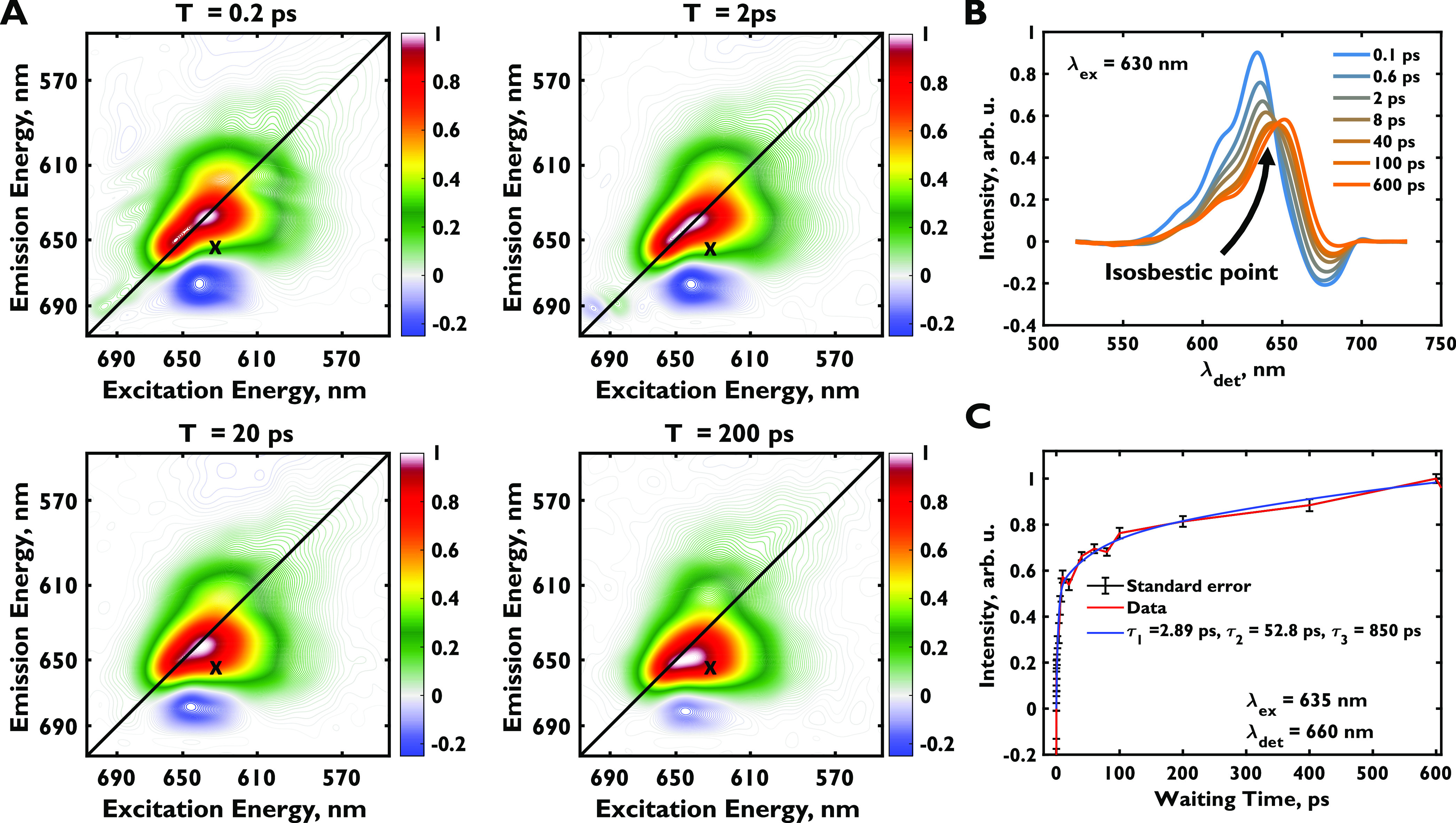
The phycobilisome is the primary light-harvesting antenna in cyanobacterial and red algal oxygenic photosynthesis. It maintains near-unity efficiency of energy transfer to reaction centers despite relying on slow exciton hopping along a relatively sparse network of highly fluorescent phycobilin chromophores. How the complex maintains this high efficiency remains unexplained. Using a two-dimensional electronic spectroscopy polarization scheme that enhances energy transfer features, we directly watch energy flow in the phycobilisome complex of Synechocystis sp. PCC 6803 from the outer phycocyanin rods to the allophycocyanin core. The observed downhill flow of energy, previously hidden within congested spectra, is faster than timescales predicted by Förster hopping along single rod chromophores. We attribute the fast, 8 ps energy transfer to interactions between rod-core linker proteins and terminal rod chromophores, which facilitate unidirectionally downhill energy flow to the core. This mechanism drives the high energy transfer efficiency in the phycobilisome and suggests that linker protein-chromophore interactions have likely evolved to shape its energetic landscape.
Conflict of interest statement
The authors declare no competing financial interest.
Figures


Similar articles
-
Linker proteins enable ultrafast excitation energy transfer in the phycobilisome antenna system of Thermosynechococcus vulcanus.Photochem Photobiol Sci. 2016 Jan;15(1):31-44. doi: 10.1039/c5pp00285k. Epub 2015 Nov 5. Photochem Photobiol Sci. 2016. PMID: 26537632
-
Annihilation of Excess Excitations along Phycocyanin Rods Precedes Downhill Flow to Allophycocyanin Cores in the Phycobilisome of Synechococcus elongatus PCC 7942.J Phys Chem B. 2022 Jan 13;126(1):23-29. doi: 10.1021/acs.jpcb.1c06509. Epub 2022 Jan 4. J Phys Chem B. 2022. PMID: 34982932 Free PMC article.
-
The phycobilisome core-membrane linkers from Synechocystis sp. PCC 6803 and red-algae assemble in the same topology.Plant J. 2021 Sep;107(5):1420-1431. doi: 10.1111/tpj.15389. Epub 2021 Jul 19. Plant J. 2021. PMID: 34171163
-
Structure of Phycobilisomes.Annu Rev Biophys. 2021 May 6;50:53-72. doi: 10.1146/annurev-biophys-062920-063657. Annu Rev Biophys. 2021. PMID: 33957054 Review.
-
Allophycocyanin and energy transfer.Biochim Biophys Acta. 2004 Jul 9;1657(2-3):73-81. doi: 10.1016/j.bbabio.2004.04.005. Biochim Biophys Acta. 2004. PMID: 15238265 Review.
Cited by
-
Intramolecular charge transfer and the function of vibronic excitons in photosynthetic light harvesting.Photosynth Res. 2024 Dec;162(2-3):139-156. doi: 10.1007/s11120-024-01095-5. Epub 2024 Apr 24. Photosynth Res. 2024. PMID: 38656684 Review.
-
Entropy is an important design principle in the photosystem II supercomplex.Proc Natl Acad Sci U S A. 2025 Mar 25;122(12):e2426331122. doi: 10.1073/pnas.2426331122. Epub 2025 Mar 19. Proc Natl Acad Sci U S A. 2025. PMID: 40106348 Free PMC article.
-
Inter-subunit energy transfer processes in a minimal plant photosystem II supercomplex.Sci Adv. 2024 Feb 23;10(8):eadh0911. doi: 10.1126/sciadv.adh0911. Epub 2024 Feb 23. Sci Adv. 2024. PMID: 38394196 Free PMC article.
References
-
- Blankenship R. E.Molecular Mechanisms of Photosynthesis, 3rd ed.; Wiley-Blackwell: Hoboken, NJ, 2021.
-
- Green B. R.; Parson W. W.. Light-Harvesting Antennas in Photosynthesis; Springer Netherlands: Dordrecht, 2003.
