. 2023 Jun 13;17(11):10327-10336.
doi: 10.1021/acsnano.3c00683. Epub 2023 May 18.

Recapitulating the Lateral Organization of Membrane Receptors at the Nanoscale

Affiliations

Recapitulating the Lateral Organization of Membrane Receptors at the Nanoscale

Seyed R Tabaei et al. ACS Nano. .

Abstract

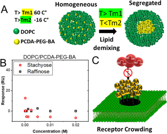
Many cell membrane functions emerge from the lateral presentation of membrane receptors. The link between the nanoscale organization of the receptors and ligand binding remains, however, mostly unclear. In this work, we applied surface molecular imprinting and utilized the phase behavior of lipid bilayers to create platforms that recapitulate the lateral organization of membrane receptors at the nanoscale. We used liposomes decorated with amphiphilic boronic acids that commonly serve as synthetic saccharide receptors and generated three lateral modes of receptor presentation─random distribution, nanoclustering, and receptor crowding─and studied their interaction with saccharides. In comparison to liposomes with randomly dispersed receptors, surface-imprinted liposomes resulted in more than a 5-fold increase in avidity. Quantifying the binding affinity and cooperativity proved that the boost was mediated by the formation of the nanoclusters rather than a local increase in the receptor concentration. In contrast, receptor crowding, despite the presence of increased local receptor concentrations, prevented multivalent oligosaccharide binding due to steric effects. The findings demonstrate the significance of nanometric aspects of receptor presentation and generation of multivalent ligands including artificial lectins for the sensitive and specific detection of glycans.

Keywords: membrane receptor; multivalent interaction; nanoclusters; receptor crowding; surface molecular imprinting.


Conflict of interest statement

The authors declare no competing financial interest.

Figures

Figure 1
Figure 1
Schematic representation of the template-guided assembly of BA clusters. (A) BA covalently and reversibly binds with 1,2- or 1,3-cis-diols to form five- or six-membered cyclic boronic esters in alkaline solution. The cyclic esters dissociate at an acidic pH. (B) Free lateral diffusion of BA receptors in the fluid lipid membrane (T > Tm), followed by (C) exposure to the target saccharide to form BA clusters upon multivalent interaction. Lipid mobility facilitates receptor (BA) recruitment by the multivalent ligand (oligosaccharide). (D) Polymerization of the matrix lipids at a temperature below the phase transition temperature (T < Tm) to “freeze” the optimum arrangement of otherwise randomly distributed BA receptors. The nanoclusters on the surface of the imprinted liposomes are stabilized by photopolymerization. The polymerization step covalently connects all monomers, thereby fixing the arrangement of receptors and preventing lipid demixing. (E) Ligand-directed rearrangement of BA.
Scheme 1
Scheme 1. Synthesis of the PCDA derivatives. (1) PCDA, (2) PCDA-BA with no PEG linker, (3) PCDA-PEG-BA, and (4) PCDA-PEG.
Figure 2
Figure 2
(A) Schematic representation of the immobilized liposomes on the SPR gold chip. Liposomes (100–200 nm) are functionalized with biotin, which is coupled to a biotinylated BSA through Neutravidin. (B) The SPR response upon injection of saccharides at different concentrations to the control (surface before liposome immobilization, dashed curves) and liposome channel (surface after liposome immobilization, solid curves). (C) The corrected SPR response corresponding to the interaction of a saccharide with BA-modified liposomes as a function of saccharide concentration. These values are the difference between the achieved constant level upon injection of saccharides at different concentrations and the control and liposome channels.
Figure 3
Figure 3
Binding curves corresponding to the interaction of tetrasaccharide stachyose and (A) nonimprinted and (B) imprinted liposomes composed of PCDA-PEG:PCDA-PEG-BA (10%). To generate imprinted liposomes, stachyose was used as the template. Binding curves of stachyose to (C) nonimprinted and (D) imprinted PCDA-PEG:PCDA-PEG-BA (25%) liposomes. (E) Schematic illustration of the effect of BA clustering on the binding behavior of the oligosaccharide. The formation of BA clusters reduces the number of binding sites accessible for binding but increases the binding strength due to multiple interactions.
Figure 4
Figure 4
Binding curves corresponding to the interaction of monosaccharide fructose with (A) nonimprinted and (B) imprinted liposomes composed of PCDA-PEG:PCDA-PEG-BA (4:1). To generate imprinted liposomes, stachyose was used as the template. Binding curves corresponding to the interaction of stachyose and fructose with liposomes composed of (C) PCDA and (D) PCDA-PEG:PCDA-BA. No significant interaction was observed. The insets show the molecular structure of PCDA and PCDA-BA with no PEG linker.
Figure 5
Figure 5
(A) Schematic depicting a fluorescence microscopy assay for detecting liposome–cell surface saccharide binding. Biotinylated 3′-sialyllactose molecules are attached to a PLL-g-PEG-biotin model surface through a biotin-Neutravidin linker. To assess the interaction of the liposomes with the surface-attached saccharides, imprinted and nonimprinted liposomes containing a trace amount of a fluorescently labeled lipid (Rhod-PE) were incubated with the surface in a microfluidic channel for a certain amount of time (10 min) and then washed to remove the unbounded liposomes. The bound liposomes were imaged by a fluorescence microscope. Representative fluorescence microscopy image of (B) imprinted and (C) nonimprinted liposomes composed of PCDA-PEG:PCDA-PEG-BA (25%) bound to the saccharide-decorated surface shown in (A). (D) No liposomes bound to the control surface with no 3′-sialyllactose. Fluorescence microscopy images of (E) imprinted and (F) nonimprinted PCDA-PEG:PCDA-PEG-BA (10%) liposomes. (G) Total number of bound liposomes per image for imprinted and nonimprinted liposomes. More representative images are presented in Supporting Figure S2.
Figure 6
Figure 6
(A) Schematic representation of the phase separation process in the liposomal membrane of a binary lipid mixture with different Tm. Lipids are homogeneously mixed at a temperature above the Tm of both lipids. Lipids undergo demixing upon cooling to ambient temperature, resulting in the formation of domains of distinct lipids. (B) SPR data corresponding to the interaction of tetrasaccharide stachyose and trisaccharide raffinose with liposomes composed of DOPC/PCDA-PEG-BA (25%). No significant interaction was observed. (C) Schematic illustration of receptor crowding and nanoclustering. When BA molecules are densely and randomly packed, they obstruct ligand binding, presumably due to the steric effect.

Similar articles

References

    1. Irvine D. J.; Purbhoo M. A.; Krogsgaard M.; Davis M. M. Direct observation of ligand recognition by T cells. Nature 2002, 419 (6909), 845–849. 10.1038/nature01076. - DOI - PubMed
    1. Davis M. M.; Boniface J. J.; Reich Z.; Lyons D. Ligand recognition by (alpha)(beta) T cell receptors. Annual review of immunology 1998, 16, 523.10.1146/annurev.immunol.16.1.523. - DOI - PubMed
    1. Schamel W. W.; Alarcón B. Organization of the resting TCR in nanoscale oligomers. Immunological reviews 2013, 251 (1), 13–20. 10.1111/imr.12019. - DOI - PubMed
    1. Molnár E.; Deswal S.; Schamel W. W. Pre-clustered TCR complexes. FEBS letters 2010, 584 (24), 4832–4837. 10.1016/j.febslet.2010.09.004. - DOI - PubMed
    1. Wolf A. A.; Jobling M. G.; Wimer-Mackin S.; Ferguson-Maltzman M.; Madara J. L.; Holmes R. K.; Lencer W. I. Ganglioside structure dictates signal transduction by cholera toxin and association with caveolae-like membrane domains in polarized epithelia. J. Cell Biol. 1998, 141 (4), 917–927. 10.1083/jcb.141.4.917. - DOI - PMC - PubMed

Publication types