. 2023 May 18;18(5):e0285735.
doi: 10.1371/journal.pone.0285735. eCollection 2023.

Presence and activity of Fibrinogen like protein 2 in platelets

Affiliations

Presence and activity of Fibrinogen like protein 2 in platelets

Izhack Cherny et al. PLoS One. .

Abstract

Background: Fibrinogen-like protein 2 (FGL2) is a serine protease capable of converting prothrombin into thrombin (i.e., prothrombinase-like activity) while bypassing the classic coagulation cascade. It has been reported to be expressed by mononuclear blood cells and endothelial cells. There are multiple reports that FGL2 supports tumor development and metastasis. However, in the blood, the origin and functional significance of FGL2 has not been established.

Objective: To determine if FGL2, a malignancy related enzyme, is present in platelets.

Methods: Peripheral blood samples were collected in K2 EDTA tubes. Blood cells and platelets were separated and thoroughly washed to produce plasma-free samples. Procoagulant activity was measured in the cell lysates using a thrombin generation test or an adjusted prothrombin time (PT) test in plasma deficient of factor X. The findings were further supported by confocal microscopy, immunoprecipitation, flow cytometry, enzyme-linked immunosorbent assays and specific inhibition assays.

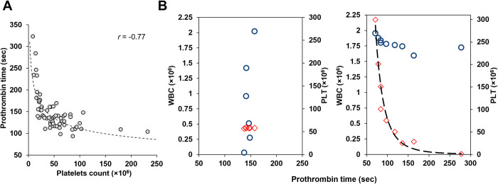
Results: FGL2 protein was readily detected in platelets. Also, despite being expressed by lymphocytes, FGL2 prothrombinase-like activity was solely detected in platelet samples, but not in white blood cell samples. Quiescent platelets were shown to contain the FGL2 protein in an active form. Upon activation, platelets secreted the active FGL2 into the milieu.

Conclusions: Active FGL2 is found in platelets. This suggests another role for the involvement of platelets in malignancies.


Conflict of interest statement

The authors have declared that no competing interests exist.

Figures

Fig 1
Fig 1. Prothrombinase activity in peripheral blood cell samples is dependent on the number of platelets in the sample.
Analysis of Prothrombin Time (clotting time shortening) in samples of peripheral blood cell from healthy individuals homogenized in factor X deficient plasma. (A) Correlation (power-regression) between clotting time and the number of platelets in samples from healthy individuals. All samples include precisely 1.5×10⁶ mononuclear cells. (B) Correlation between Prothrombin time and counts of platelets (◇) or white blood cells (○). Platelets and white blood cells from a single donor were purified separately and then combined to generate different combinations. Left panel B, constant platelets count (58 × 106) and increasing amounts of white blood cells (0.03–2×10⁶); Right panel B, similar white blood cells count (1.6–2×10⁶) and increasing amounts of platelets (0–1.5 × 10⁶).
Fig 2
Fig 2. The detected procoagulant activity in platelets is prothrombinase.
(A) A total of 2×10⁸ platelets were homogenized in factor-X deficient plasma (FXDP), or factor-II deficient plasma (FIIDP). Clotting was induced using the prothrombin time (PT) method. The time of clotting was recorded and compared to the time of clotting generated by samples of FXDP, or FIIDP only (without platelets). While both FIIDP samples elicit clotting after 97 seconds, regardless of the presence of platelets (Δ = 0±8 seconds), FXDP clotting time in the presence or absence of platelets was 109 and 397 seconds, respectively (Δ = 288±20 seconds). The measurements were performed in quadruplicates. Error bars represent standard deviation. (B) Twelve sample pairs of 108 platelets were homogenized in FXDP versus FVDP and tested for prothrombin time activity. The correlation between prothrombin time generated by the samples in FVDP versus FXDP was weak (r = 0.38) and insignificant (p = 0.198). (C) Mean PT induced by PC3 wild type cells (228±19 sec) or FGL2 overexpressing cells (179±14 sec) homogenized in FXDP. Experiments were performed in triplicate. Error bars represent standard deviation (p-value = 0.01). FGL2 overexpression was confirmed by western blot analysis. β-actin was used as a control.
Fig 3
Fig 3. FGL2 is present in platelets.
(A) Immunoprecipitation of FGL2 and FX from plasma-free peripheral blood samples. Immunoprecipitating antibodies and matched normal IgG subclasses are indicated. Mock represents nonspecific binding to protein A/G-agarose beads. Expected FX and FGL2 protein sizes are indicated by arrows. Only FGL2 was precipitated. (B) FGL2 and FX quantification using Enzyme-linked immunosorbent assays. Protein concentrations were determined in purified platelet samples (109 platelets per sample, n = 11 healthy individuals), in plasma samples (n = 4 healthy individuals), in a normal pool plasma and in an FX-deficient plasma. Bars represent mean concentration. Error bars represent the standard deviation. (C) Confocal immunodetection of FGL2 or FX in freshly prepared non-stimulated platelets, assisted by FITC-conjugated secondary antibodies. Platelet membranes were fluorolabeled with CellMask deep red reagent. Background fluorescence (i.e., FITC-conjugated antibody only) is presented as well.
Fig 4
Fig 4. Flow cytometry detection of FGL2 in platelets and lymphocytes.
The fluorescence intensity of IgG control-alexafluor 488 (Red) or FGL2-alexafluor 488 (Green) antibodies was detected in gated platelet using log SSC/FSC/CD41+ or in gated lymphocyte using CD45+/SSC plot following cell permeabilization (A) or intact cells (B).
Fig 5
Fig 5. Validation of FGL2 activity in platelets.
(A) inhibition of FGL2 activity by Anti-NPG-12 antibody. Platelets samples from peripheral blood were homogenized in FXDP (Stago, France) and incubated with increasing concentrations of Rabbit anti-NPG-12 antibody capable of specifically inhibiting FGL2 prothrombinase-like activity. Normal Rabbit IgG was used as a control. The resulting inhibition is expressed in percentages relative to the measured activity in the absence of antibody. Experiment was performed by three independent repeats (B) Inhibition of FX activity by rivaroxaban. Platelets samples from peripheral blood were homogenized in FXDP (IL, Italy) and incubated with increasing concentrations of rivaroxaban. FXDP and Normal pooled plasma were used as controls. The resulting inhibition is expressed in percentages of FX activity (log scale). The experiment was performed as two independent repeats. Error bars represent standard deviation.
Fig 6
Fig 6. Platelets secrete active FGL2 upon activation.
Identical and equal suspensions of 1×10⁸ platelets were treated with indicated agonists to stimulate platelets activation or maintain quiescence. The soluble fractions, containing the secreted proteins, were separated from the platelets fraction by centrifugation. (A) FGL2 was immunodetected by western blot in the supernatant of the activated platelets. (B) Activity was measured in the soluble fractions (containing the secreted proteins) by the prothrombin time assay. Clotting time shortening (represented by a red circle) was evident in the soluble fractions of the activated platelets, but not of the quiescent platelets. The column bars represent the corresponding activity, expressed as a percentage relative to the maximum activity obtained following complete homogenization of the sample (left bar). The experiment was performed in triplicates. Error bars represent standard deviation.

Similar articles

References

    1. Chan CWY, Chan MWC, Liu M, Fung L, Cole EH, Leibowitz JL, et al.. Kinetic analysis of a unique direct prothrombinase, fgl2, and identification of a serine residue critical for the prothrombinase activity. J Immunol. 2002;168(10):5170–5177. doi: 10.4049/jimmunol.168.10.5170 - DOI - PubMed
    1. Yang G, Hooper WC. Physiological functions and clinical implications of fibrinogen-like 2: A review. World J Clin Infect Dis. 2013;3(3):37–46. doi: 10.5495/wjcid.v3.i3.37 - DOI - PMC - PubMed
    1. Yu J, Li J, Shen J, Du F, Wu X, Li M, et al.. The role of Fibrinogen-like proteins in Cancer. Int J Biol Sci. 2021;17(4):1079–1087. doi: 10.7150/ijbs.56748 - DOI - PMC - PubMed
    1. Marazzi S, Blum S, Hartmann R, Gundersen D, Schreyer M, Argraves S, et al.. Characterization of human fibroleukin, a fibrinogen-like protein secreted by T lymphocytes. J Immunol. 1998;161(1):138–147. - PubMed
    1. Liu H, Shalev I, Manuel J, He W, Leung E, Crookshank J, et al.. The FGL2-FcgammaRIIB pathway: a novel mechanism leading to immunosuppression. Eur J Immunol. 2008; 38(11):3114–3126. doi: 10.1002/eji.200838338 - DOI - PubMed

Publication types