. 2023 Jun 16;18(6):1315-1323.
doi: 10.1021/acschembio.3c00033. Epub 2023 May 18.

Anthracyclines React with Apurinic/Apyrimidinic Sites in DNA

Anthracyclines React with Apurinic/Apyrimidinic Sites in DNA

Medjda Bellamri et al. ACS Chem Biol. .

Abstract

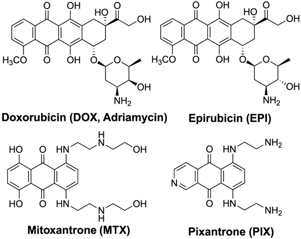
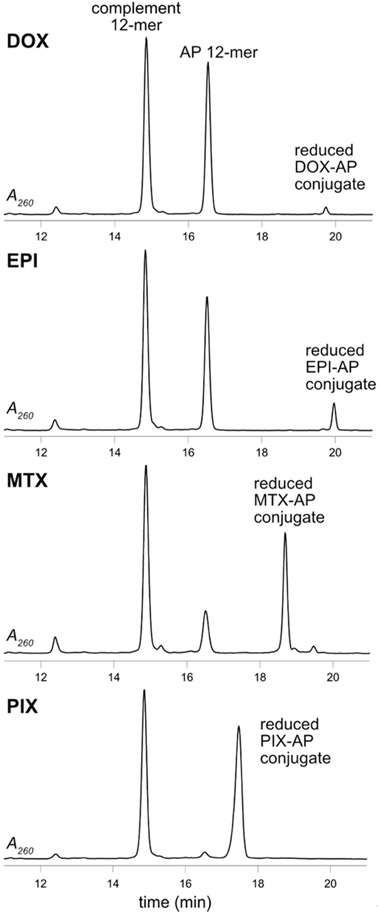
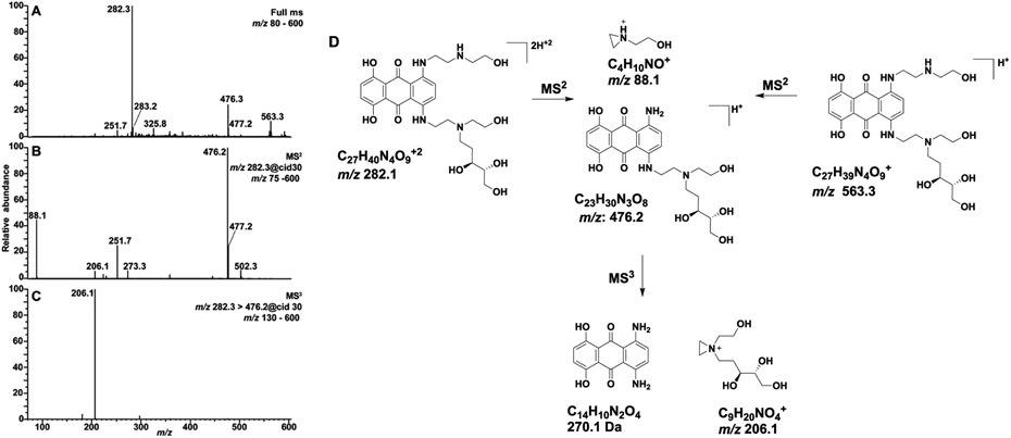
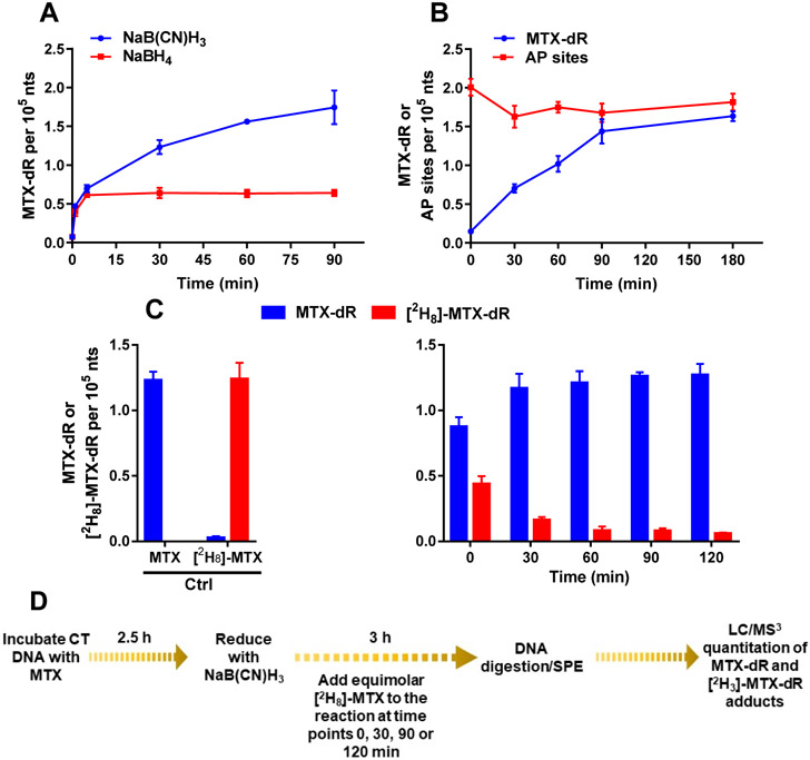
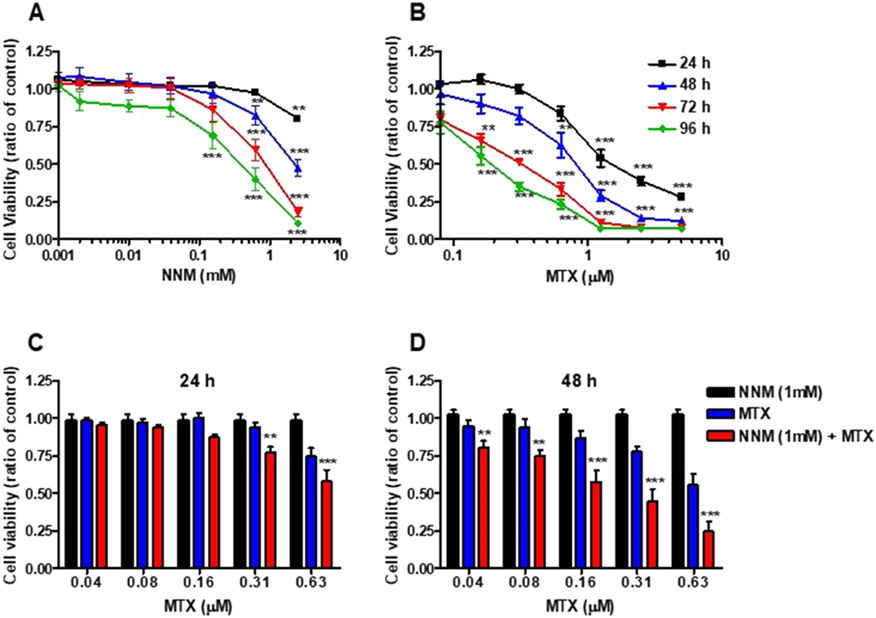
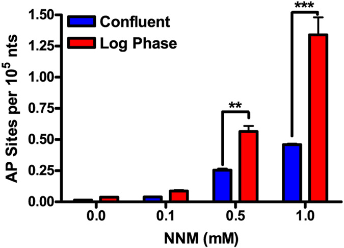
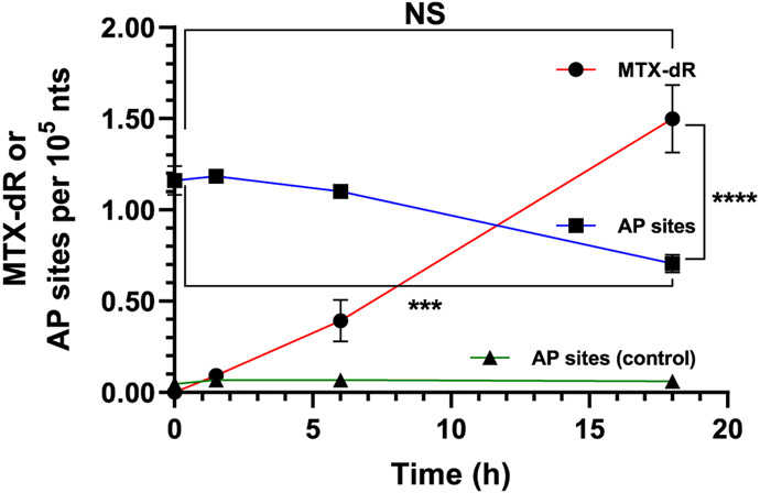
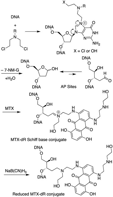
The combination of doxorubicin (Adriamycin) and cyclophosphamide, referred to as AC chemotherapy, is commonly used for the clinical treatment of breast and other cancers. Both agents target DNA with cyclophosphamide causing alkylation damage and doxorubicin stabilizing the topoisomerase II-DNA complex. We hypothesize a new mechanism of action whereby both agents work in concert. DNA alkylating agents, such as nitrogen mustards, increase the number of apurinic/apyrimidinic (AP) sites through deglycosylation of labile alkylated bases. Herein, we demonstrate that anthracyclines with aldehyde-reactive primary and secondary amines form covalent Schiff base adducts with AP sites in a 12-mer DNA duplex, calf thymus DNA, and MDA-MB-231 human breast cancer cells treated with nor-nitrogen mustard and the anthracycline mitoxantrone. The anthracycline-AP site conjugates are characterized and quantified by mass spectrometry after NaB(CN)H3 or NaBH4 reduction of the Schiff base. If stable, the anthracycline-AP site conjugates represent bulky adducts that may block DNA replication and contribute to the cytotoxic mechanism of therapies involving combinations of anthracyclines and DNA alkylating agents.


Figures

Figure 1.
Figure 1.
Clinically used anthracycline antitumor agents.
Figure 2.
Figure 2.
HPLC-UV chromatograms of the reaction of the AP-containing 12-mer duplex with DOX, EPI, MTX, and PIX reduced with NaB(CN)H3 analyzed after 6 h.
Figure 3.
Figure 3.
(A) Full-scan mass spectrum of the synthetic MTX-dR reduced conjugate showing the singly protonated [M + H]+ at m/z 563.3 and the doubly protonated [M + 2H]+2 ion at m/z 282.3. (B) The MS2 scan stage mass spectrum of m/z 282.3 and (C) MS3 spectrum of the m/z 476.2 product ion are reported. (D) Proposed fragmentation mechanisms of the major ions.
Figure 4.
Figure 4.
Kinetics of MTX-dR Schiff base formation in CT DNA reduced with (A) NaB(CN)H3 or NaBH4. (B) AP sites in CT DNA derivatized with PMOA and MTX-dR Schiff base reduced with NaB(CN)H3. (C) The displacement of MTX-dR in CT DNA with [2H8]-MTX during reduction with NaB(CN)H3. (D) The displacement experiment scheme with [2H8]-MTX. Adduct levels are reported as the mean ± SD (n = 3 replicates for MTX-dR and n = 4 replicates for AP sites derivatized with PMOA). The levels of PMOA-dR and reduced MTX-dR at 180 min were not significantly different.
Figure 5.
Figure 5.
Cytotoxicity of NNM and MTX in MDA-MB-231 cells. Cells were treated with (A) NNM [0.001–2.5 mM], (B) MTX [0.04–5.0 μM], or solvent control for up to 96 h. Cell viability was measured by the MTT assay every 24 h. Cytotoxicity of NNM and MTX cotreatment in MDA-MB-231 cells (C) after 24 h, and (D) after 48 h. Cells were treated with NNM [1 mM], in the presence of MTX [0.04–0.63 μM], or solvent control. Cell viability was measured by the MTT assay after 24 or 48 h. The data are compiled from three different experiments [mean ± SD]. (two-way ANOVA, Benferroni's multiple comparisons post test, NNM + MTX vs MTX, *P < 0.05; **P < 0.01, ***P < 0.005).
Figure 6.
Figure 6.
Concentration-dependent increase in AP site formation in MDA-MB-231 cells. After 24 h of treatment with NNM [0.1–1 mM], AP site levels were measured following derivatization with PMOA. The data are compiled from three different experiments [mean ± SD]. Two-way ANOVA, confluence vs log phase (***P < 0.01, ***P < 0.005).
Figure 7.
Figure 7.
MDA-MB-231 cells cotreated with 1 mM NNM and 0.6 μM MTX for 24 h. Reduced MTX-dR levels formed over 18 h in the isolated nuclei treated with NaB(CN)H3. AP site levels and stability in the nuclei of untreated and NNM-treated cells were measured at the same time points as done for MTX-dR reduction. Adduct levels are reported as the mean ± SEM (n = 4 independent replicates, ***P < 0.005, ****P < 0.0001, NS, not significant).
Scheme 1.
Scheme 1.. Depurination N7-Nitrogen Mustard dG Adducts Leading to an AP Site and Subsequent Conjugation by MTXa
aThe MTX conjugate is reduced with NaB(CN)H3 and detected by LC/MS.

References

    1. Jones SE; Durie BG; Salmon SE Combination chemotherapy with adriamycin and cyclophosphamide for advanced breast cancer. Cancer 1975, 36, 90–97. - PubMed
    1. Martins-Teixeira MB; Carvalho I Antitumour anthracyclines: Progress and perspectives. ChemMedChem 2020, 15, 933–948. - PubMed
    1. Povirk LF; Shuker DE DNA damage and mutagenesis induced by nitrogen mustards. Mutat. Res 1994, 318, 205–226. - PubMed
    1. Chen H; Cui Z; Hejazi L; Yao L; Walmsley SJ; Rizzo CJ; Turesky RJ Kinetics of DNA adducts and abasic site formation in tissues of mice treated with a nitrogen mustard. Chem. Res. Toxicol 2020, 33, 988–998. - PMC - PubMed
    1. Chen H; Yao L; Brown C; Rizzo CJ; Turesky RJ Quantitation of apurinic/apyrimidinic sites in isolated DNA and in mammalian tissue with a reduced level of artifacts. Anal. Chem 2019, 91, 7403–7410. - PMC - PubMed

Publication types