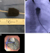
Treatment of Esophageal Perforation: Endoscopic Vacuum-Assisted Closure
- PMID: 37200717
- PMCID: PMC10187847
- DOI: 10.1097/PG9.0000000000000314
Treatment of Esophageal Perforation: Endoscopic Vacuum-Assisted Closure
Abstract
Surgical repair of type C esophageal atresia (EA) with distal tracheoesophageal fistula (TEF) is complicated by an anastomotic leak in 10%-30% of cases with associated morbidity. A novel procedure in the pediatric population, endoscopic vacuum-assisted closure (EVAC), accelerates the healing of esophageal leaks by using the effects of VAC therapy, including fluid removal and stimulation of granulation tissue formation. We report 2 additional cases of chronic esophageal leak treated with EVAC in EA patients. The first is a patient with a previously repaired type C EA/TEF and left congenital diaphragmatic hernia complicated by an infected diaphragmatic hernia patch erosion into the esophagus and colon. Additionally, we discuss a second case using EVAC for early anastomotic leak following type C EA/TEF repair in a patient who was later found to have a distal congenital esophageal stricture.
Keywords: anastomosis; hernia; pediatrics; sponge; tracheoesophageal fistula.
Copyright © 2023 The Author(s). Published by Wolters Kluwer Health, Inc. on behalf of the European Society for Pediatric Gastroenterology, Hepatology, and Nutrition and the North American Society for Pediatric Gastroenterology, Hepatology, and Nutrition.
Conflict of interest statement
The authors report no conflicts of interest.
Figures


References
-
- Manfredi MA, Jennings RW, Anjum MW, et al. Externally removable stents in the treatment of benign recalcitrant strictures and esophageal perforations in pediatric patients with esophageal atresia. Gastrointest Endosc. 2014;80:246–252. - PubMed
-
- Cameron JL, Keiffer RF, Hendrix TR, et al. Selective nonoperative management of contained intrathoracic esophageal disruptions. Ann Thorac Surg. 1979;27:404–408. - PubMed
-
- Huang C, Leavitt T, Bayer LR, et al. Effect of negative pressure wound therapy on wound healing. Curr Probl Surg. 2014;51:301–331. - PubMed
-
- Smithers CJ, Hamilton TE, Manfredi MA, et al. Categorization and repair of recurrent and acquired tracheoesophageal fistulae occurring after esophageal atresia repair. J Pediatr Surg. 2017;52:424–430. - PubMed
-
- Nirula R. Esophageal perforation. Surg Clin North Am. 2014;94:35–41. - PubMed
Publication types
LinkOut - more resources
Full Text Sources

