. 2023 May 2:10:1105678.
doi: 10.3389/fmolb.2023.1105678. eCollection 2023.

In silico analysis of the profilaggrin sequence indicates alterations in the stability, degradation route, and intracellular protein fate in filaggrin null mutation carriers

Affiliations

In silico analysis of the profilaggrin sequence indicates alterations in the stability, degradation route, and intracellular protein fate in filaggrin null mutation carriers

Argho Aninda Paul et al. Front Mol Biosci. .

Abstract

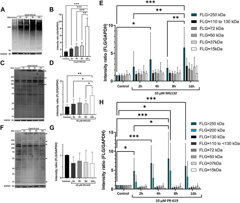
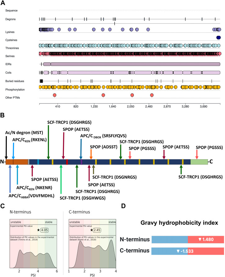
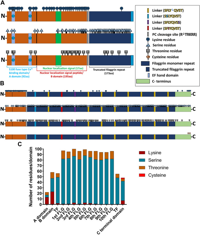
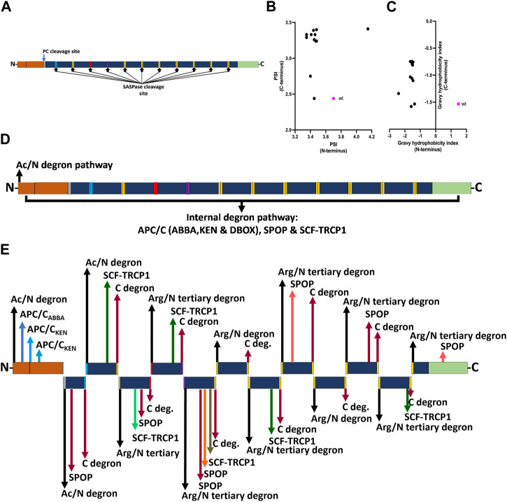
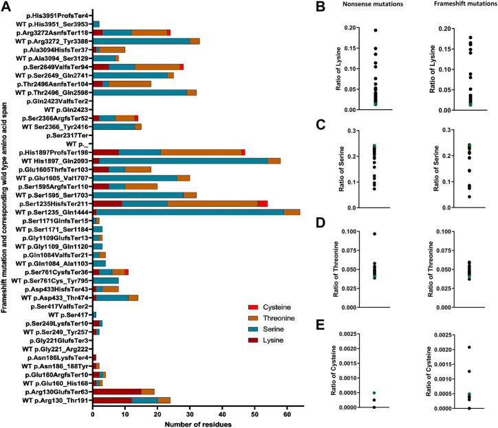
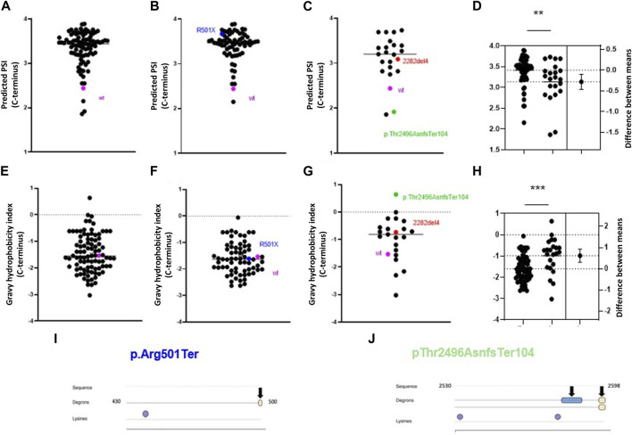
Background: Loss of function mutation in FLG is the major genetic risk factor for atopic dermatitis (AD) and other allergic manifestations. Presently, little is known about the cellular turnover and stability of profilaggrin, the protein encoded by FLG. Since ubiquitination directly regulates the cellular fate of numerous proteins, their degradation and trafficking, this process could influence the concentration of filaggrin in the skin. Objective: To determine the elements mediating the interaction of profilaggrin with the ubiquitin-proteasome system (i.e., degron motifs and ubiquitination sites), the features responsible for its stability, and the effect of nonsense and frameshift mutations on profilaggrin turnover. Methods: The effect of inhibition of proteasome and deubiquitinases on the level and modifications of profilaggrin and processed products was assessed by immunoblotting. Wild-type profilaggrin sequence and its mutated variants were analysed in silico using the DEGRONOPEDIA and Clustal Omega tool. Results: Inhibition of proteasome and deubiquitinases stabilizes profilaggrin and its high molecular weight of presumably ubiquitinated derivatives. In silico analysis of the sequence determined that profilaggrin contains 18 known degron motifs as well as multiple canonical and non-canonical ubiquitination-prone residues. FLG mutations generate products with increased stability scores, altered usage of the ubiquitination marks, and the frequent appearance of novel degrons, including those promoting C-terminus-mediated degradation routes. Conclusion: The proteasome is involved in the turnover of profilaggrin, which contains multiple degrons and ubiquitination-prone residues. FLG mutations alter those key elements, affecting the degradation routes and the mutated products' stability.

Keywords: atopic dermatitis; degron; filaggrin; proteasome; ubiquitination.


Conflict of interest statement

The authors declare that the research was conducted in the absence of any commercial or financial relationships that could be construed as a potential conflict of interest.

Figures

FIGURE 1
FIGURE 1
UPS is involved in degradation of profilaggrin. (A) Western blot with anti-ubiquitin in the keratinocytes upon treatment with proteasome inhibitor MG132; (B) Intensity ratio of ubiquitinated protein bands after proteasome inhibition for 2 h, 4 h, 8 h and 16 h; one-way ANOVA followed with Tukey’s multiple comparison test. (C) Western blot with anti-filaggrin in keratinocytes upon 2 h, 4 h, 8 h and 16 h treatment of proteasome inhibitor MG132, arrows indicate accumulation of undegraded profilaggrin in proteasome inhibition sample. (D) Intensity ratio of all filaggrin bands detected in different timepoint treatment of proteasome inhibitor MG132; one-way ANOVA followed by Tukey’s multiple comparison test; (E) Intensity ratio of different FLG bands upon MG132 treatment; two-way ANOVA followed by Šidák’s multiple comparison test; (F) Western blot with anti-filaggrin in keratinocytes upon 2 h, 4 h, 8 h and 16 h treatment of deubiquitinase inhibitor PR-619, Top two arrows and top box indicates accumulation of higher molecular weight filaggrin bands in the deubiquitinase inhibited samples whereas, deubiquitinase inhibition leads to depletion or disappearance of filaggrin bands of approximately 130, 50 and 15 kDa pointed with bottom box and arrows. (G) Intensity ratio of all filaggrin bands detected in different timepoint treatment of deubiquitinase inhibitor PR-619; one-way ANOVA followed by Tukey’s multiple comparison test; (H) Intensity ratio of different FLG bands upon PR-619 treatment; two-way ANOVA followed by Šidák’s multiple comparison test; error bar stands for + SD and p-value < 0.001(***); p-value ≤0.002(**); p-value ≤ 0.033(*); (n = 4).
FIGURE 2
FIGURE 2
Location of degron motifs, ubiquitination-prone residues, and terminal stability of profilaggrin. (A) Illustration showing location of degron, ubiquitin-conjugating amino acid residues, intrinsic disorder region, phosphorylation sites and other post-translational modification sites in the profilaggrin protein. (B) Location of degron motif sequence and degron type in the profilaggrin wild-type sequence (legend as of Figure 3); (C) protein stability index of the N- and C- terminus of the profilaggrin; (D) Gravy hydrophobicity index of profilaggrin N- and C terminus.
FIGURE 3
FIGURE 3
Distribution of ubiquitin (Ub)-conjugating residues in the profilaggrin wildtype sequence. (A) Illustration of N terminus of FLG. The N terminal of profilaggrin consists of a S100 fused type Ca++ Binding domain (92 aa) also known as A domain followed by nuclear localization signal peptide (195 aa) also known as B domain and a truncated filaggrin repeat (173 aa). Truncated filaggrin repeat is flanked with a PC cleavage site which links it with B domain and SASPase cleavage site which links it with rest of the filaggrin. Like all the other S100 domain filaggrin S100 domain consists of the Ca++ binding domain followed by a unique nuclear localization signal. (B) Illustration of the wild-type profilaggrin protein. (C) Number of Ub-conjugating amino acids in different domains of wild type profilaggrin.
FIGURE 4
FIGURE 4
Effect of profilaggrin processing on the stability and degron content. (A) PC and SASPase clevage sites in the profilaggrin sequence. (B) terminal protein stability index of newly generated SASPase cleaved FLG products in comparison to the wild-type FLG terminus. (C) terminal Gravy hydrophobicity index of newly generated SASPase cleaved FLG products in comparison to the wild-type FLG terminus. (D) Proposed degradation routes of wild-type profilaggrin. (E) Proposed degradation routes of PC- and SASPase-cleaved profilaggrin products (legends as of Figure 3).
FIGURE 5
FIGURE 5
Altered content of ubiquitin-conjugating residues in FLG mutation products. (A) Number of ubiquitin-conjugating residues in the frameshift products in comparison to the corresponding wild-type same length span. (B) Ratio of lysine in the nonsense and frameshift mutant protein. (C) Ratio of serine (Ser) in the nonsense and frameshift mutant protein. (D) Ratio of threonine (Thr) in the nonsense and frameshift mutant protein. (E) Ratio of cysteine in the nonsense and frameshift mutant protein.
FIGURE 6
FIGURE 6
Alterations in profilaggrin stability in FLG null mutation carriers. (A–D) C-terminal PSI of recurrent pathogenic frameshift (C), nonsense mutation (B) and combined (A) here wild type is shown as pink and two most common European mutations 2282del4 and R500X are marked in red and blue; (D) estimation plot and comparison between PSI scores; Student’s t-test; p < 0.01 (**); (E–H) C terminal Gravy hydrophobicity index of recurrent pathogenic frameshift (G), nonsense mutation (F) and combined (E) here wild type is shown as pink and two most renowned mutation 2282del4 and R500X are marked in red and blue; (H) estimation plot and comparison between PSI scores; Student’s t-test; p < 0.001 (***); (I) Location of C-terminal degron in p.Arg501Ter and Lysine residue. (J) Location of C terminal degrons and lysine residues in the p.Thr2496AsnfsTer104.

References

    1. Abbas T., Dutta A. (2009). P21 in cancer: Intricate networks and multiple activities. Nat. Rev. Cancer 9, 400–414. 10.1038/nrc2657 - DOI - PMC - PubMed
    1. Aho S., Harding C. R., Lee J. M., Meldrum H., Bosko C. A. (2012). Regulatory role for the profilaggrin N-terminal domain in epidermal homeostasis. J. Investigative Dermatology 132, 2376–2385. 10.1038/jid.2012.174 - DOI - PubMed
    1. Aksnes H., Drazic A., Marie M., Arnesen T. (2016). First things first: Vital protein marks by N-terminal acetyltransferases. Trends Biochem. Sci. 41, 746–760. 10.1016/j.tibs.2016.07.005 - DOI - PubMed
    1. Backendorf C., Hohl D. (1992). A common origin for cornified envelope proteins? Nat. Genet. 2, 91. 10.1038/ng1092-91 - DOI - PubMed
    1. Barker J. N. W. N., Palmer C. N. A., Zhao Y., Liao H., Hull P. R., Lee S. P., et al. (2007). Null mutations in the filaggrin gene (FLG) determine major susceptibility to early-onset atopic dermatitis that persists into adulthood. J. Invest. Dermatol 127, 564–567. 10.1038/sj.jid.5700587 - DOI - PubMed