. 2023 Apr 29;14(2):1064-1076.
doi: 10.21037/jgo-23-198. Epub 2023 Apr 23.

Exosomal miR-125b-5p derived from cancer-associated fibroblasts promotes the growth, migration, and invasion of pancreatic cancer cells by decreasing adenomatous polyposis coli (APC) expression

Affiliations

Exosomal miR-125b-5p derived from cancer-associated fibroblasts promotes the growth, migration, and invasion of pancreatic cancer cells by decreasing adenomatous polyposis coli (APC) expression

Yuting Guo et al. J Gastrointest Oncol. .

Abstract

Background: A significant desmoplastic response, particularly in the fibroblasts, is a characteristic of pancreatic ductal adenocarcinoma (PDAC). Increasing evidence has shown that cancer-associated fibroblasts (CAFs) assist tumor development, invasion, and metastasis in PDAC. However, CAFs-derived molecular determinants that regulate the molecular mechanisms of PDAC have not been fully characterized.

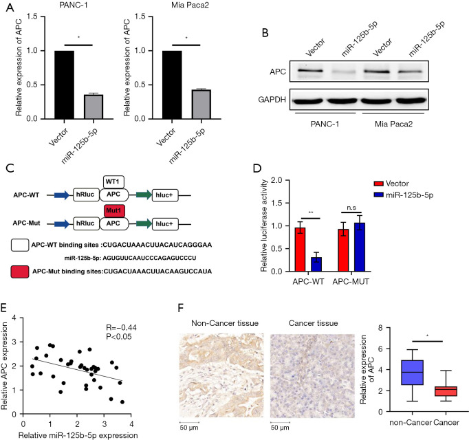
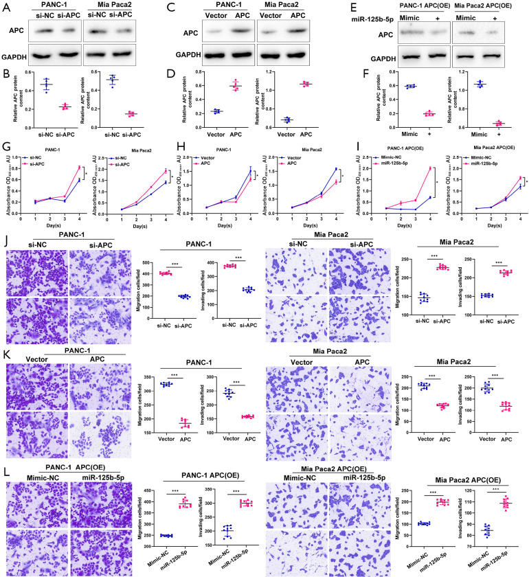
Methods: The expression of microRNA 125b-5p (miR-125b-5p) in Pancreas Cancer (PC) tissue and para-cancerous normal tissue was examined using Polymerase Chain Reaction (PCR). Cell counting kit-8 (CCK8), wound healing, and transwell experiments were utilized to assess the effect of miR-125b-5p. Using a cell luciferase activity test and bioinformatics, it was demonstrated that miR-125b-5p may bind to the 3'-untranslated region (3'-UTR) of the adenomatous polyposis coli (APC), thereby limiting the progression of pancreatic cancer.

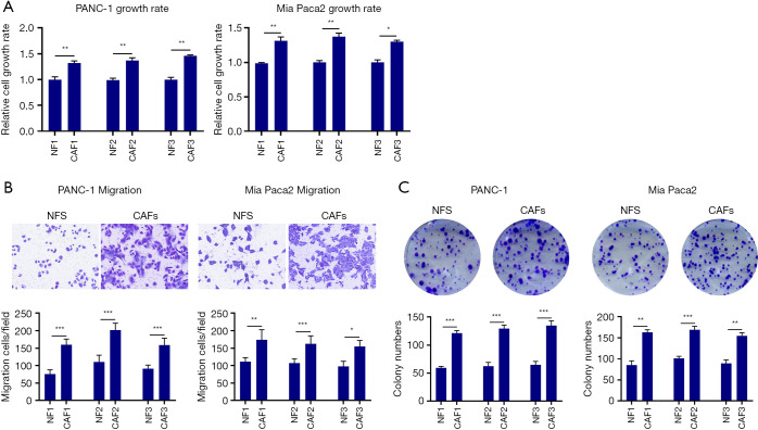
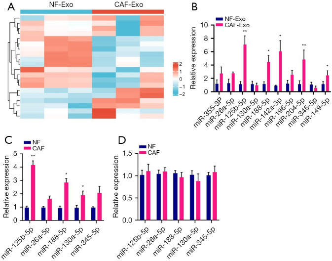
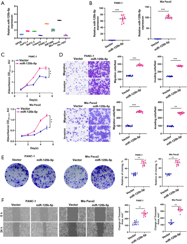
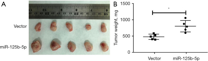
Results: PDAC cells are prompted to proliferate, undergo the epithelial-mesenchymal transition (EMT), and spread. Importantly, CAFs release exosomes into PDAC cells, which significantly increase the level of miR-125b-5p in those cells. Meanwhile, pancreatic cancer cell lines and PDAC tissues have considerably higher miR-125b-5p expression. MiR-125b-5p's elevated expression mechanically suppresses the expression of APC and accelerates the spread of pancreatic cancer.

Conclusions: Exosomes released by CAFs promote PDAC growth, invasion, and metastasis. Exosomal miR-125b-5p inhibition offers an alternate strategy for combating the basic malady of PDAC.

Keywords: Pancreatic cancer; adenomatous polyposis coli (APC); cancer-associated fibroblasts (CAFs); exosomes; miR-125b-5p.


Conflict of interest statement

Conflicts of Interest: All authors have completed the ICMJE uniform disclosure form (available at https://jgo.amegroups.com/article/view/10.21037/jgo-23-198/coif). The authors have no conflicts of interest to declare.

Figures

Figure 1
Figure 1
Pancreatic cancer cells’ proliferative and invasive metastatic abilities are enhanced by CAF. (A) CCK8 assay analysis of the proliferative viability of the pancreatic cancer cells when co-cultured with CAF cells; (B) a Transwell experiment was used to detect the invasion and migration of pancreatic cancer cells co-cultured with CAF cells. Images were taken at a 400× zoom using a Zeiss lens; (C) plate cloning assay analysis of the formation ability of pancreatic cancer cell colony co-cultured with CAF cells. (B,C) Stained with 0.1% crystal violet. *P<0.05, **P<0.01 and ***P<0.001. NF, normal fib; CAF, cancer-associated fibroblast; CCK8, cell counting kit 8.
Figure 2
Figure 2
miR-125b-5p was highly expressed in CAF exosomes. (A) Transcriptome sequencing analysis of miRNA expression in normal fibroblast and tumor-associated fibroblast exosomes; (B,C) qRT-PCR analysis of differentially expressed miRNAs in normal fibroblast and tumor-associated fibroblast exosomes; (D) qRT-PCR analysis of differentially expressed pre-miRNAs in normal fibroblasts and tumor-associated fibroblasts. *P<0.05, **P<0.01. NF, Normal Fib; CAF, cancer-associated fibroblast; qRT-PCR, quantitative real-time polymerase chain reaction.
Figure 3
Figure 3
miR-125b-5p promotes pancreatic cancer proliferation, migration, and invasion. (A) qRT-PCR detection of miR-125b-5p expression in normal ductal epithelial cells of the pancreas and pancreatic cancer cells; (B) qRT-PCR detection of miR-125b-5p expression differences between the Vector and miR-125b-5p overexpression groups; (C) CCK8 assay was performed to detect changes in the proliferation ability of pancreatic cancer cells in the Vector and miR-125b-5p overexpression groups; (D) Transwell assay to detect changes in the proliferation ability of pancreatic cancer cells in the Vector and miR-125b-5p overexpression groups. Images were taken at a 400× zoom using a Zeiss lens; (E) plate cloning assay to detect changes in the proliferation ability of pancreatic cancer cells in the Vector and miR-125b-5p overexpression groups; (F) cell scratch assay was performed to detect changes in the proliferation ability of pancreatic cancer cells in the Vector and miR-125b-5p overexpression groups. Images were taken at a 400× zoom using a Zeiss lens. (D,E) Stained with 0.1% crystal violet. **P<0.01 and ***P<0.001. qRT-PCR, quantitative real-time polymerase chain reaction; CCK8, cell counting kit 8.
Figure 4
Figure 4
miR-125b-5p directly targets APC. (A) qRT-PCR assay was used to analyze the difference in APC expression in the Vector and miR-125b-5p overexpression groups; (B) Western blot assay was used to analyze the difference in APC expression in the Vector and miR-125b-5p overexpression groups; (C) schematic diagram of vector construction for wild-type and mutant APC luciferase; (D) dual luciferase to verify the binding ability of miR-125b-5p to APC; (E) qRT-PCR analysis of the correlation between miR-125b-5p and APC expression levels; (F) immunohistochemical analysis of APC expression levels in paraneoplastic and cancerous tissues. The sections were subjected to DAB reaction. Images were taken at a 400× zoom using a Zeiss lens *P<0.05, **P<0.01, n.s, no significance. APC, adenomatous polyposis coli; WT, wild-type; MUT, mutant; qRT-PCR, quantitative real-time polymerase chain reaction.
Figure 5
Figure 5
miR-125b-5p-targeted APC promotes pancreatic cancer progression. (A,B) Immunoblotting assay to detect the difference of APC expression in the control and APC-interfering groups; (C,D) immunoblotting assay to detect the difference of APC expression in the control and overexpression groups; (E,F) analysis of the effect of miR-125b-5p on APC expression by immunoblotting in APC-overexpressing cells; (G) CCK8 assay to analyze the proliferation ability of cells in the control and APC-interfering groups; (H) CCK8 assay to analyze the cell proliferation ability in the control and APC overexpression groups; (I) CCK8 assay analysis of the effect of miR-125b-5p on cell proliferation capacity in APC-overexpressed cells; (J) Transwell assay to analyze cell migration ability in the control and APC-interfering groups; (K) Transwell assay to analyze cell migration ability in the control and APC-overexpression groups; (L) analysis of the effect of miR-125b-5p on cell migration ability by Transwell assay in APC-overexpressed cells. Stained with 0.1% crystal violet. Images were taken at a 400× zoom using a Zeiss lens. *P<0.05, ***P<0.001 APC, adenomatous polyposis coli; GAPDH, glyceraldehyde-3-phosphate dehydrogenase; OE, overexpression; CCK8, cell counting kit 8.
Figure 6
Figure 6
miR-125b-5p inhibits pancreatic cancer cell proliferation in vivo. (A) Nude mouse tumorigenic assay to analyze the effect of miR-125b-5p on the proliferative capacity of pancreatic cancer cells in vivo; (B) analysis of tumor weights in the control and miR-125b-5p-overexpression groups. *P<0.05.

Similar articles

Cited by

References

    1. Yang J, Li Y, Han X, et al. The antitumor effect of the novel agent MCL/ACT001 in pancreatic ductal adenocarcinoma. J Cancer Res Clin Oncol 2022. 10.1007/s00432-022-04542-9 - DOI - PMC - PubMed
    1. Du L, Wang-Gillam A. Trends in Neoadjuvant Approaches in Pancreatic Cancer. J Natl Compr Canc Netw 2017;15:1070-7. 10.6004/jnccn.2017.0134 - DOI - PubMed
    1. Bejarano L, Jordao MJC, Joyce JA. Therapeutic Targeting of the Tumor Microenvironment. Cancer Discov 2021;11:933-59. 10.1158/2159-8290.CD-20-1808 - DOI - PubMed
    1. Jin MZ, Jin WL. The updated landscape of tumor microenvironment and drug repurposing. Signal Transduct Target Ther 2020;5:166. 10.1038/s41392-020-00280-x - DOI - PMC - PubMed
    1. Mao X, Xu J, Wang W, et al. Crosstalk between cancer-associated fibroblasts and immune cells in the tumor microenvironment: new findings and future perspectives. Mol Cancer 2021;20:131. 10.1186/s12943-021-01428-1 - DOI - PMC - PubMed