Review
. 2023 Apr 29;14(2):1114-1130.
doi: 10.21037/jgo-22-1034. Epub 2023 Mar 29.

Advances in pre-treatment evaluation of pancreatic ductal adenocarcinoma: a narrative review

Affiliations
Review

Advances in pre-treatment evaluation of pancreatic ductal adenocarcinoma: a narrative review

Michele Fiore et al. J Gastrointest Oncol. .

Abstract

Background and objective: Despite advances in the multidisciplinary management of pancreatic cancer, overall prognosis remains poor, due to early progression of the disease. There is a need to also take action in staging, to make it increasingly accurate and complete, to define the setting of the therapeutic strategy. This review was planned to update the current status of pre-treatment evaluation for pancreatic cancer.

Methods: We conducted an extensive review, including relevant articles dealing with traditional imaging, functional imaging and minimally invasive surgical procedures before treatment for pancreatic cancer. We searched articles written in English only. Data in the PubMed database, published in the period between January 2000 and January 2022, were retrieved. Prospective observational studies, retrospective analyses and meta-analyses were reviewed and analysed.

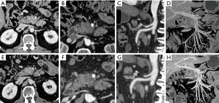
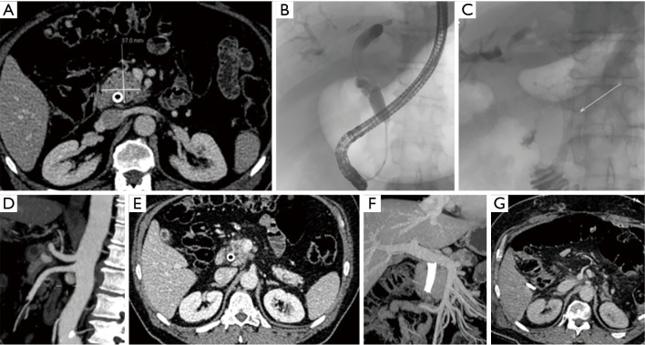
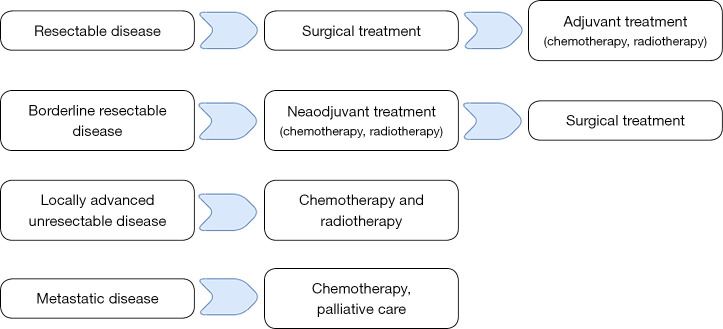
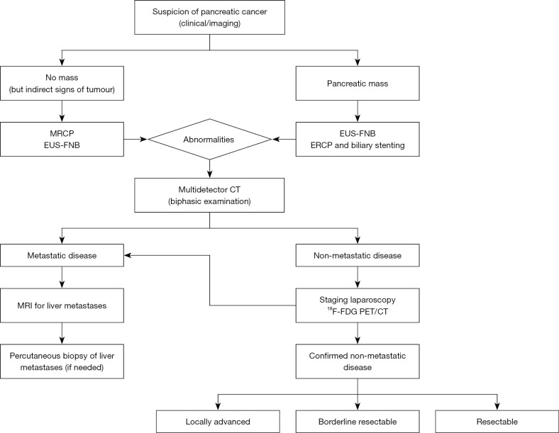
Key content and findings: Each imaging modality (endoscopic ultrasonography, endoscopic retrograde cholangiopancreatography, computed tomography, positron emission tomography/computed tomography, staging laparoscopy) has its own diagnostic advantages and limitations. The sensitivity, specificity and accuracy for each image set are reported. Data that support the increasing role of neoadjuvant therapy (radiotherapy and chemotherapy) and the meaning of a patient-tailored treatment selection, based on tumour staging, are also discussed.

Conclusions: A multimodal pre-treatment workup should be searched as it improves staging accuracy, orienting patients with resectable tumors towards surgery, optimizing patient selection with locally advanced tumors to neoadjuvant or definite therapy and avoiding surgical resection or curative radiotherapy in those with metastatic disease.

Keywords: Pancreatic cancer; imaging; laparoscopy; staging.


Conflict of interest statement

Conflicts of Interest: All authors have completed the ICMJE uniform disclosure form (available at https://jgo.amegroups.com/article/view/10.21037/jgo-22-1034/coif). The series “Pancreas Surgery” was commissioned by the editorial office without any funding or sponsorship. AC served as the unpaid Guest Editor of the series. The authors have no other conflicts of interest to declare.

Figures

Figure 1
Figure 1
CT scan displaying a (A) biopsy-proven adenocarcinoma of the pancreatic head (52 mm × 32 mm); (B) radiologic evidence of >180° tumor interface to superior mesenteric artery, (C) extending for about 4 cm, and (D) signs of venous occlusion affecting the distal III of the superior mesenteric vein, for about 2.5 cm, suggesting the presence of an unresectable lesion. CT scan performed after 3 months from neoadjuvant chemoradiation showing (E) reduction (43 mm × 28 mm) of the lesion, (F) the presence of <180° tumor interface to superior mesenteric artery (arrow), extending for about 34 mm (G) and (H) the decrease of the extension of venous occlusion, feature of borderline resectable cancer. CT, computed tomography.
Figure 2
Figure 2
CT scan displaying a (A) biopsy-proven pancreatic adenocarcinoma (45 mm × 37 mm), which required the endoscopic positioning of a metallic stent due to the biliary tree dilatation. (B) ERCP fluoroscopic images highlight the biliary tree dilatation and (C) the biliary detention after the stent positioning (arrow). (D) No evidence of extension of tumor to the SMA and CA, with (E,F) radiologic evidence of <180° tumor interface to superior mesenteric vein, all features of borderline resectable cancer. (G) CT scan was performed after total spleno-pancreasectomy, with no evidence of residual disease. CT, computed tomography; ERCP, endoscopic retrograde cholangiopancreatography; SMA, superior mesenteric artery; CA, celiac axis.
Figure 3
Figure 3
Treatments for patients with pancreatic cancer at different stages.
Figure 4
Figure 4
Multimodality pre-treatment workup for pancreatic cancer staging. Multidetector CT with a biphasic examination is the first-line imaging for diagnosis and tumour staging. MRCP can display abnormalities of the entire pancreatic and bile duct, in presence of indirect signs of tumour. No other imaging study is required in metastatic pancreatic cancer, with the exception of MRI of the liver to detect small metastatic lesions, not visible on standard CT in about 10% of cases. A EUS-FNB is mandatory in presence of a pancreatic mass before treatment. ERCP can be considered in patients with non resectable pancreatic cancer, who is a candidate for first line chemotherapy, or in those patients with a malignant biliary duct obstruction who need to be treated with a neoadjuvant therapy before surgery. In non-metastatic pancreatic cancer, after initial CT assessment both laparoscopy and 18F-FDG PET/CT can potentially add information to the staging workup. The potential incremental benefit of laparoscopy for staging is due to the identification of occult abdominal and peritoneal metastases, undetectable by imaging techniques. PET/CT scan has the capacity to detect lymph nodes and distant metastases, particularly in borderline resectable and locally advanced pancreatic cancer, as well as in resectable disease. MRCP, magnetic resonance cholangiopancreatography; ERCP, endoscopic retrograde cholangiopancreatography; EUS-FNB, endoscopic ultrasonography-fine needle biopsy; CT, computed tomography; MRI, magnetic resonance imaging; 18F-FDG, 18F-Fluorodeoxyglucose; PET, positron emission tomography.

Similar articles

Cited by

References

    1. Siegel RL, Miller KD, Fuchs HE, et al. Cancer statistics, 2022. CA Cancer J Clin 2022;72:7-33. 10.3322/caac.21708 - DOI - PubMed
    1. Rahib L, Smith BD, Aizenberg R, et al. Projecting cancer incidence and deaths to 2030: the unexpected burden of thyroid, liver, and pancreas cancers in the United States. Cancer Res 2014;74:2913-21. 10.1158/0008-5472.CAN-14-0155 - DOI - PubMed
    1. McGuigan A, Kelly P, Turkington RC, et al. Pancreatic cancer: A review of clinical diagnosis, epidemiology, treatment and outcomes. World J Gastroenterol 2018;24:4846-61. 10.3748/wjg.v24.i43.4846 - DOI - PMC - PubMed
    1. National Cancer Institute. Cancer Stat Facts: Pancreatic Cancer. Surveillance, Epidemiology and End Results Program. 2019.
    1. Vincent A, Herman J, Schulick R, et al. Pancreatic cancer. Lancet 2011;378:607-20. 10.1016/S0140-6736(10)62307-0 - DOI - PMC - PubMed