

A Projection-Domain Low-Count Quantitative SPECT Method for α-Particle-Emitting Radiopharmaceutical Therapy
- PMID: 37201111
- PMCID: PMC10191330
- DOI: 10.1109/trpms.2022.3175435
A Projection-Domain Low-Count Quantitative SPECT Method for α-Particle-Emitting Radiopharmaceutical Therapy
Abstract
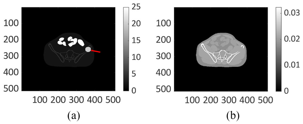
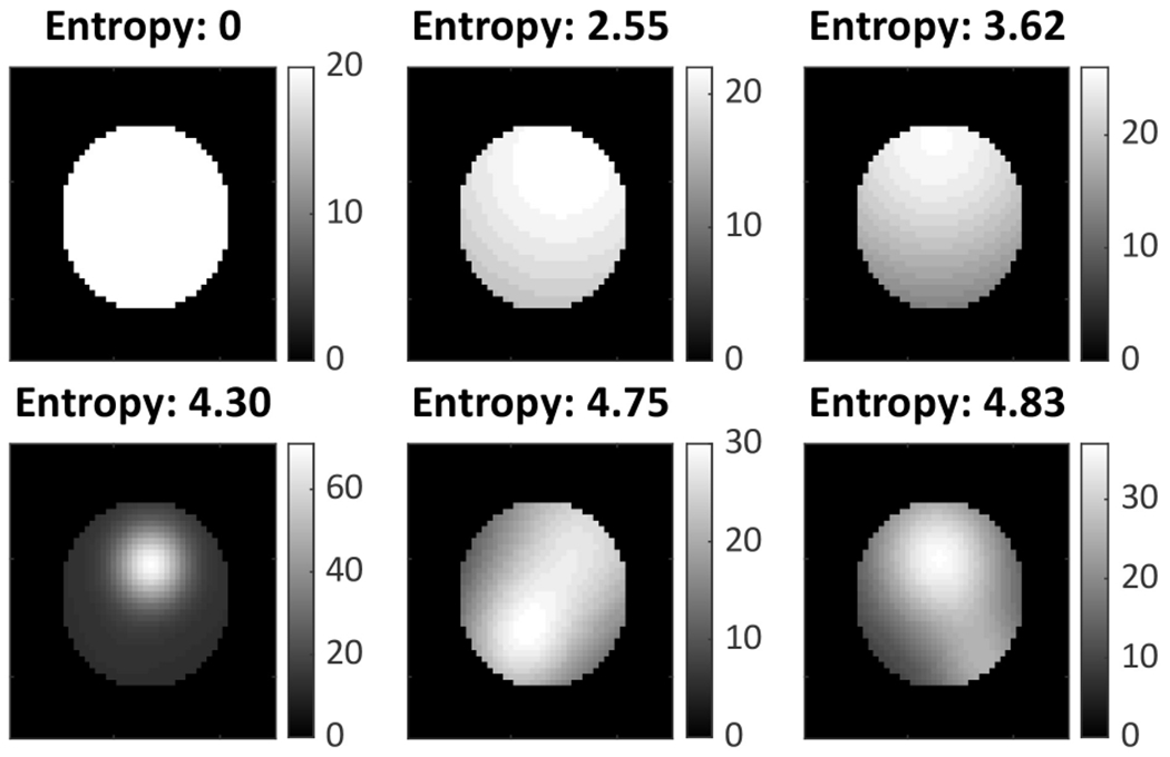
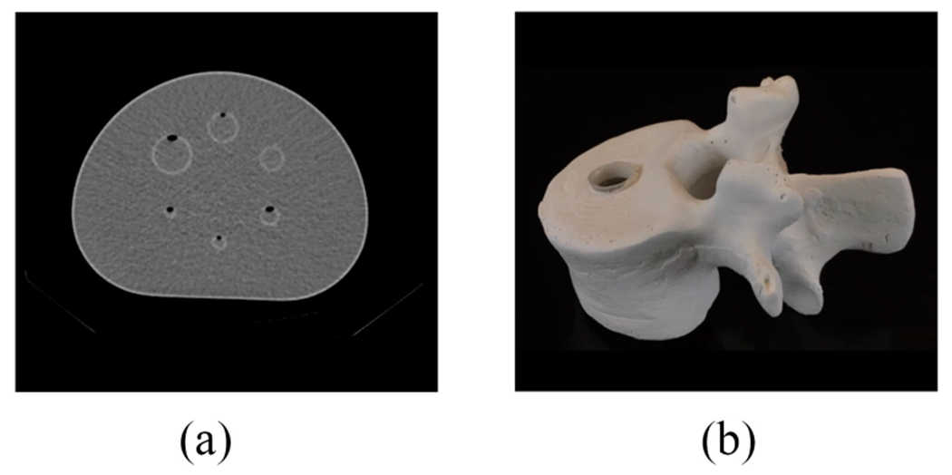
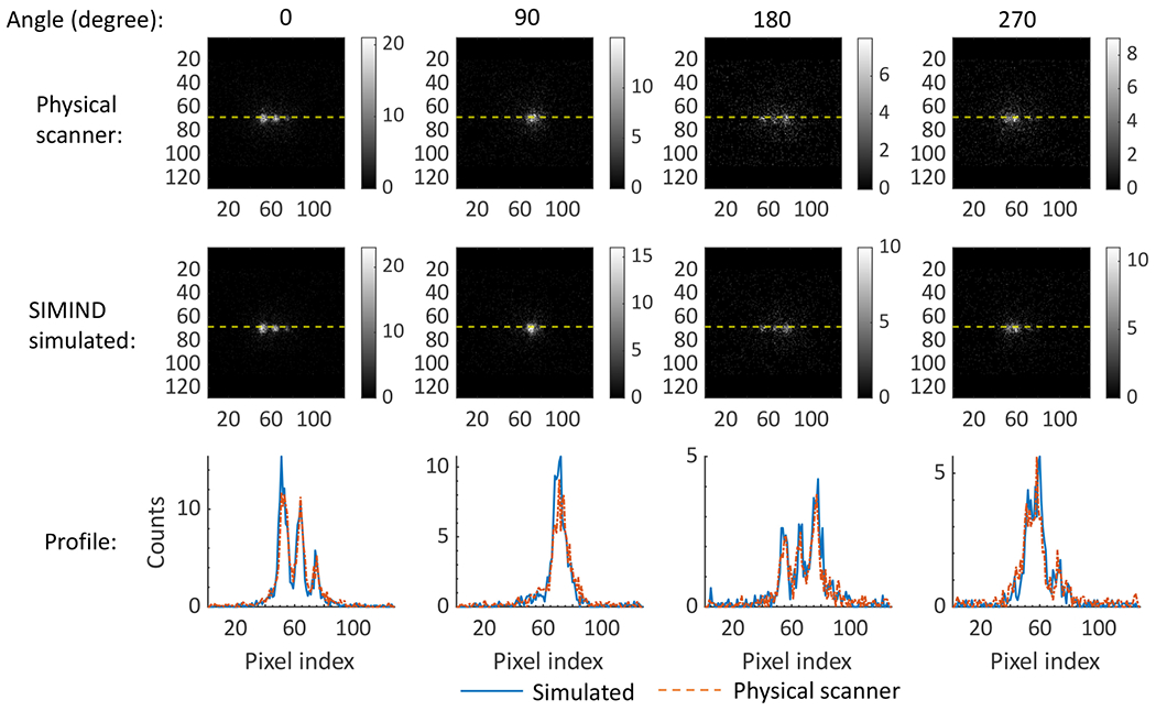
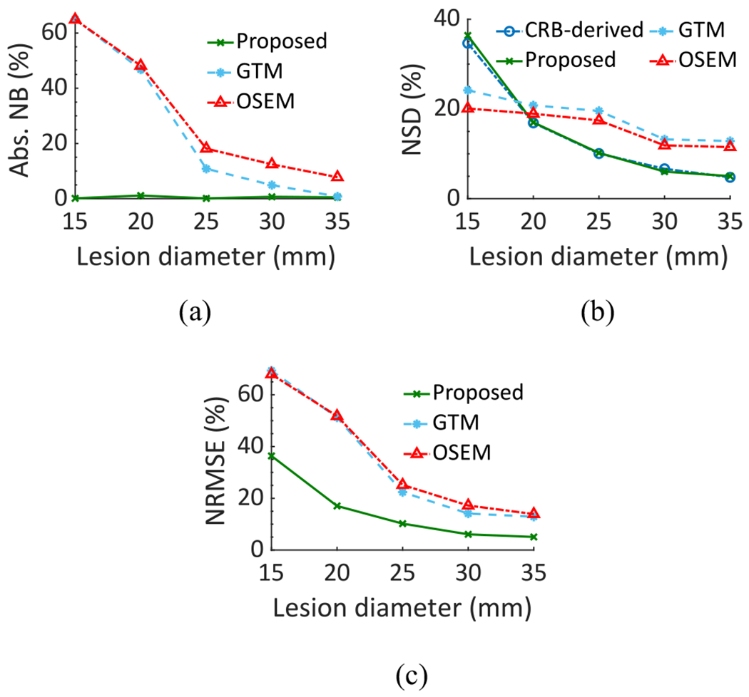
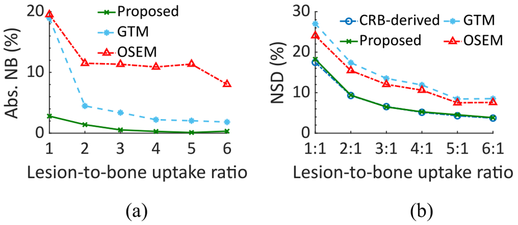
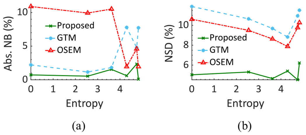
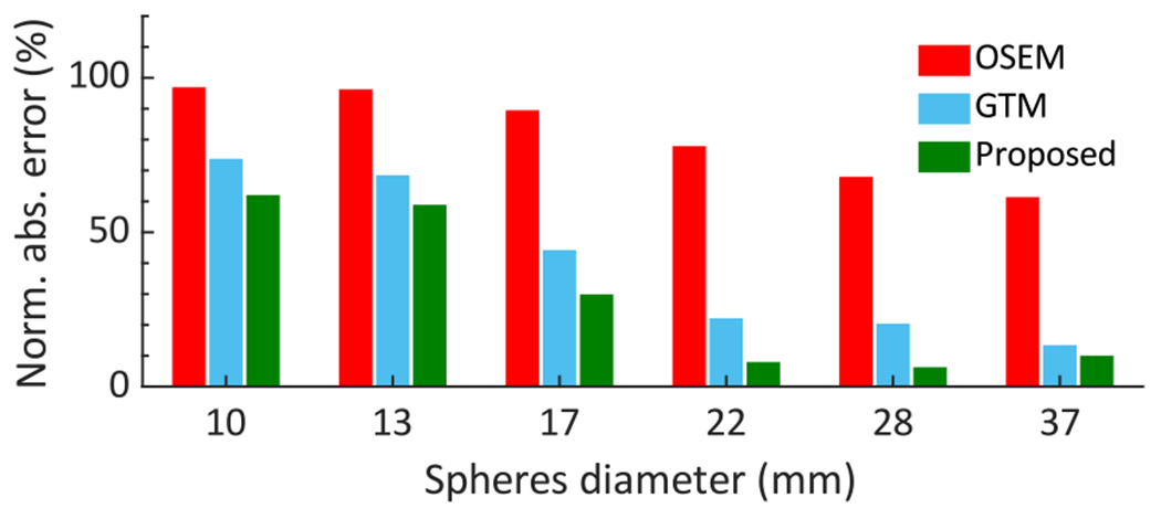
Single-photon emission-computed tomography (SPECT) provides a mechanism to estimate regional isotope uptake in lesions and at-risk organs after administration of α-particle-emitting radiopharmaceutical therapies (α-RPTs). However, this estimation task is challenging due to the complex emission spectra, the very low number of detected counts (~20 times lower than in conventional SPECT), the impact of stray-radiation-related noise at these low counts, and the multiple image-degrading processes in SPECT. The conventional reconstruction-based quantification methods are observed to be erroneous for α-RPT SPECT. To address these challenges, we developed a low-count quantitative SPECT (LC-QSPECT) method that directly estimates the regional activity uptake from the projection data (obviating the reconstruction step), compensates for stray-radiation-related noise, and accounts for the radioisotope and SPECT physics, including the isotope spectra, scatter, attenuation, and collimator-detector response, using a Monte Carlo-based approach. The method was validated in the context of 3-D SPECT with 223Ra, a commonly used radionuclide for α-RPT. Validation was performed using both realistic simulation studies, including a virtual clinical trial, and synthetic and 3-D-printed anthropomorphic physical-phantom studies. Across all studies, the LC-QSPECT method yielded reliable regional-uptake estimates and outperformed the conventional ordered subset expectation-maximization (OSEM)-based reconstruction and geometric transfer matrix (GTM)-based post-reconstruction partial-volume compensation methods. Furthermore, the method yielded reliable uptake across different lesion sizes, contrasts, and different levels of intralesion heterogeneity. Additionally, the variance of the estimated uptake approached the Cramér-Rao bound-defined theoretical limit. In conclusion, the proposed LC-QSPECT method demonstrated the ability to perform reliable quantification for α-RPT SPECT.
Keywords: Radium-223; low counts; quantitative single-photon emission-computed tomography (SPECT); regional quantification; α-particle therapies.
Figures
References
-
- Kluetz PG et al. , “Radium Ra 223 dichloride injection: U.S. Food and Drug Administration drug approval summary,” Clin. Cancer Res, vol. 20, no. 1, pp. 9–14, 2014. - PubMed
-
- Kratochwil C et al. , “Targeted α-therapy of metastatic castration-resistant prostate cancer with 225Ac-PSMA-617: Dosimetry estimate and empiric dose finding,” J. Nucl. Med, vol. 58, no. 10, pp. 1624–1631, 2017. - PubMed
-
- Jurcic JG et al. , “Targeted α particle immunotherapy for myeloid leukemia,” Blood, vol. 100, no. 4, pp. 1233–1239, 2002. - PubMed
Grants and funding
LinkOut - more resources
Full Text Sources