. 2023 May 2:13:1113401.
doi: 10.3389/fcimb.2023.1113401. eCollection 2023.

Inhibitory effects of vaginal Lactobacilli on C andida albicans growth, hyphal formation, biofilm development, and epithelial cell adhesion

Affiliations

Inhibitory effects of vaginal Lactobacilli on C andida albicans growth, hyphal formation, biofilm development, and epithelial cell adhesion

Tomonori Takano et al. Front Cell Infect Microbiol. .

Abstract

Introduction: Antifungal agents are not always efficient in resolving vulvovaginal candidiasis (VVC), a common genital infection caused by the overgrowth of Candida spp., including Candida albicans, or in preventing recurrent infections. Although lactobacilli (which are dominant microorganisms constituting healthy human vaginal microbiota) are important barriers against VVC, the Lactobacillus metabolite concentration needed to suppress VVC is unknown.

Methods: We quantitatively evaluated Lactobacillus metabolite concentrations to determine their effect on Candida spp., including 27 vaginal strains of Lactobacillus crispatus, L. jensenii, L. gasseri, Lacticaseibacillus rhamnosus, and Limosilactobacillus vaginalis, with inhibitory abilities against biofilms of C. albicans clinical isolates.

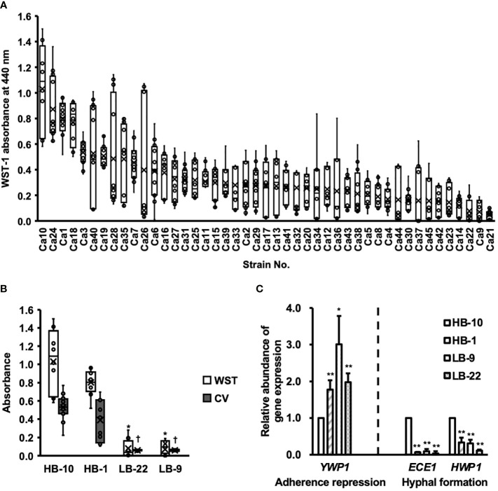
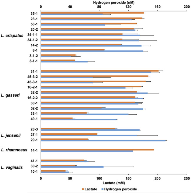
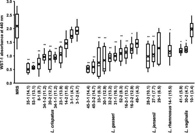
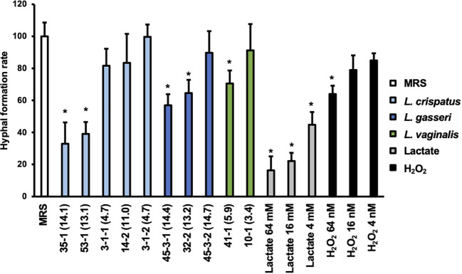
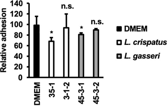
Results: Lactobacillus culture supernatants suppressed viable fungi by approximately 24%-92% relative to preformed C. albicans biofilms; however, their suppression differed among strains and not species. A moderate negative correlation was found between Lactobacillus lactate production and biofilm formation, but no correlation was observed between hydrogen peroxide production and biofilm formation. Both lactate and hydrogen peroxide were required to suppress C. albicans planktonic cell growth. Lactobacillus strains that significantly inhibited biofilm formation in culture supernatant also inhibited C. albicans adhesion to epithelial cells in an actual live bacterial adhesion competition test.

Discussion: Healthy human microflora and their metabolites may play important roles in the development of new antifungal agent against C. albicans-induced VVC.

Keywords: Candida albicans; Lactobacillus species; biofilm; cell adhesion; probiotics.


Conflict of interest statement

HaK, SE, AM, KO, and MT are employees of Miyarisan Pharmaceutical Co., Ltd.; however, they have no conflicts of interest to declare regarding this study. The remaining authors declare that the research was conducted in the absence of any commercial or financial relationships that could be construed as a potential conflict of interest.

Figures

Figure 1
Figure 1
Clinical isolates of Candida albicans with different abilities to form biofilms. CV, crystal violet; WST, water-soluble tetrazolium salts. (A) The biofilm formation of 45 clinical isolates of C albicans were used to measure WST-1 and are exhibited by the box whisker plots. Box plot shows the median (horizontal thick blank line), mean (cross), and first and third quartiles (box). (B) Biofilm formation by different C albicans strains was estimated using both WST-1 reduction and CV staining. *p < 0.05 by U-test compared with WST-1 absorbance of C albicans HB-10. p < 0.05 by U-test compared with crystal violet (CV) absorbance of C albicans HB-10. (C) Relative quantitation of genes associated with adherence repression (YWP1), or hyphal formation (HWP1 and ECE1) normalized to the β-actin gene. The C albicans HB-10 strain was used as the reference to depict the difference among the four C albicans clinical isolates. Bars represent the standard deviation from the mean values. *p < 0.05 and **p < 0.01 by U-test.
Figure 2
Figure 2
Lactate and hydrogen peroxide production by 27 Lactobacillus clinical isolates. Twenty-seven Lactobacillus clinical isolates were cultured in de Man, Rogosa, and Sharpe (MRS) broth for 72 h, and cell-free culture supernatants were collected. Lactate level was measured quantitatively by high-performance liquid chromatography (HPLC), and hydrogen peroxide was measured quantitatively using a hydrogen peroxide assay kit. Data are represented by the mean across the three replicates.
Figure 3
Figure 3
Metabolic activity of the biofilm of C. albicans HB-10 treated with culture supernatants of 27 different Lactobacillus clinical isolates. The x-axis indicates the strain number and lactic acid concentration (mM). The burden of viable cells of preformed biofilm treated after culture supernatants of 27 different Lactobacillus clinical isolates was measured using the WST-1 reduction reaction. MRS broth was used as the control. WST, water-soluble tetrazolium salts; MRS, de Man, Rogosa, and Sharpe. Box plot shows the median (horizontal thick blank line), mean (cross), and first and third quartiles (box). Bars represent the standard deviation from the mean values. *p < 0.05 and **p < 0.01 by U-test.
Figure 4
Figure 4
Hyphal formation rate of C. albicans HB-10 strain treated with Lactobacillus culture supernatants, lactate, or hydrogen peroxide. The x-axis indicates the strain number and lactic acid concentration (mM) or lactate concentration or hydrogen peroxide concentration. Relative hyphal formation in C. albicans HB-10 treated with culture supernatants of 10 different Lactobacillus clinical isolates, lactate, or hydrogen peroxide. Bars represent the standard deviation from the mean values. MRS broth was used as the control. MRS, de Man, Rogosa, and Sharpe. *p < 0.05 by U-test.
Figure 5
Figure 5
Adhesion of C. albicans HB-10 strain to HeLa cells according to the presence or absence of lactobacilli. Relative adherence of the C. albicans HB-10 strain to HeLa cells pretreated with DMEM or different lactobacilli. Bars represent the standard deviation from the mean values. *p < 0.05 by U-test. DMEM, Dulbecco’s modified Eagle’s medium. n.s., not significant.

References

    1. Aarti C., Khusro A., Varghese R., Arasu M. V., Agastian P., Al-Dhabi N. A., et al. . (2018). In vitro investigation on probiotic, anti-candida, and antibiofilm properties of Lactobacillus pentosus strain LAP1. Arch. Oral. Biol. 89, 99–106. doi: 10.1016/J.ARCHORALBIO.2018.02.014 - DOI - PubMed
    1. Alves R., Mota S., Silva S., F Rodrigues C., P Brown A. J., Henriques M., et al. . (2017). The carboxylic acid transporters Jen1 and Jen2 affect the architecture and fluconazole susceptibility of Candida albicans biofilm in the presence of lactate. Biofouling 33, 943–954. doi: 10.1080/08927014.2017.1392514 - DOI - PubMed
    1. Berman J., Sudbery P. E. (2002). Candida albicans: a molecular revolution built on lessons from budding yeast. Nat. Rev. Genet. 3, 918–931. doi: 10.1038/nrg948 - DOI - PubMed
    1. Bizerra F. C., Nakamura C. V., de Poersch C., Estivalet Svidzinski T. I., Borsato Quesada R. M., Goldenberg S., et al. . (2008). Characteristics of biofilm formation by Candida tropicalis and antifungal resistance. FEMS Yeast Res. 8, 442–450. doi: 10.1111/J.1567-1364.2007.00347.X - DOI - PubMed
    1. Bongomin F., Gago S., Oladele R. O., Denning D. W. (2017). Global and multi-national prevalence of fungal diseases-estimate precision. J. Fungi (Basel) 3. doi: 10.3390/JOF3040057 - DOI - PMC - PubMed

LinkOut - more resources