. 2023 Jul:71:50-62.
doi: 10.1016/j.mito.2023.05.004. Epub 2023 May 16.

The cyclic lipopeptide micafungin induces rupture of isolated mitochondria by reprograming the mitochondrial inner membrane anion channel

Affiliations

The cyclic lipopeptide micafungin induces rupture of isolated mitochondria by reprograming the mitochondrial inner membrane anion channel

Jonathan Hosler et al. Mitochondrion. 2023 Jul.

Abstract

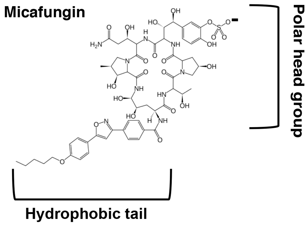
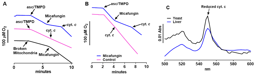
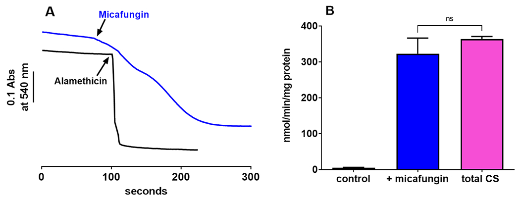
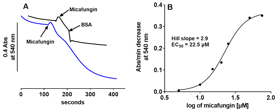
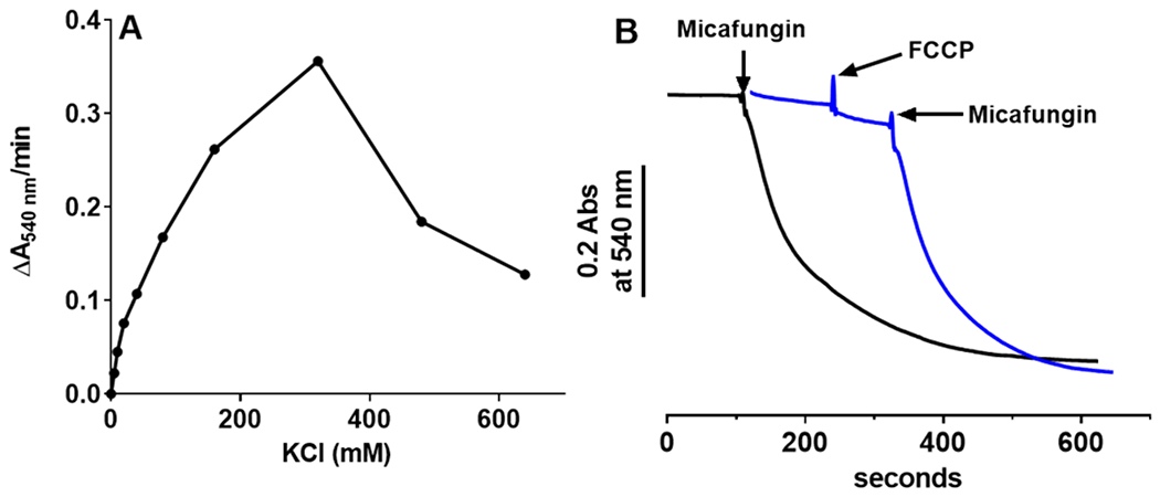
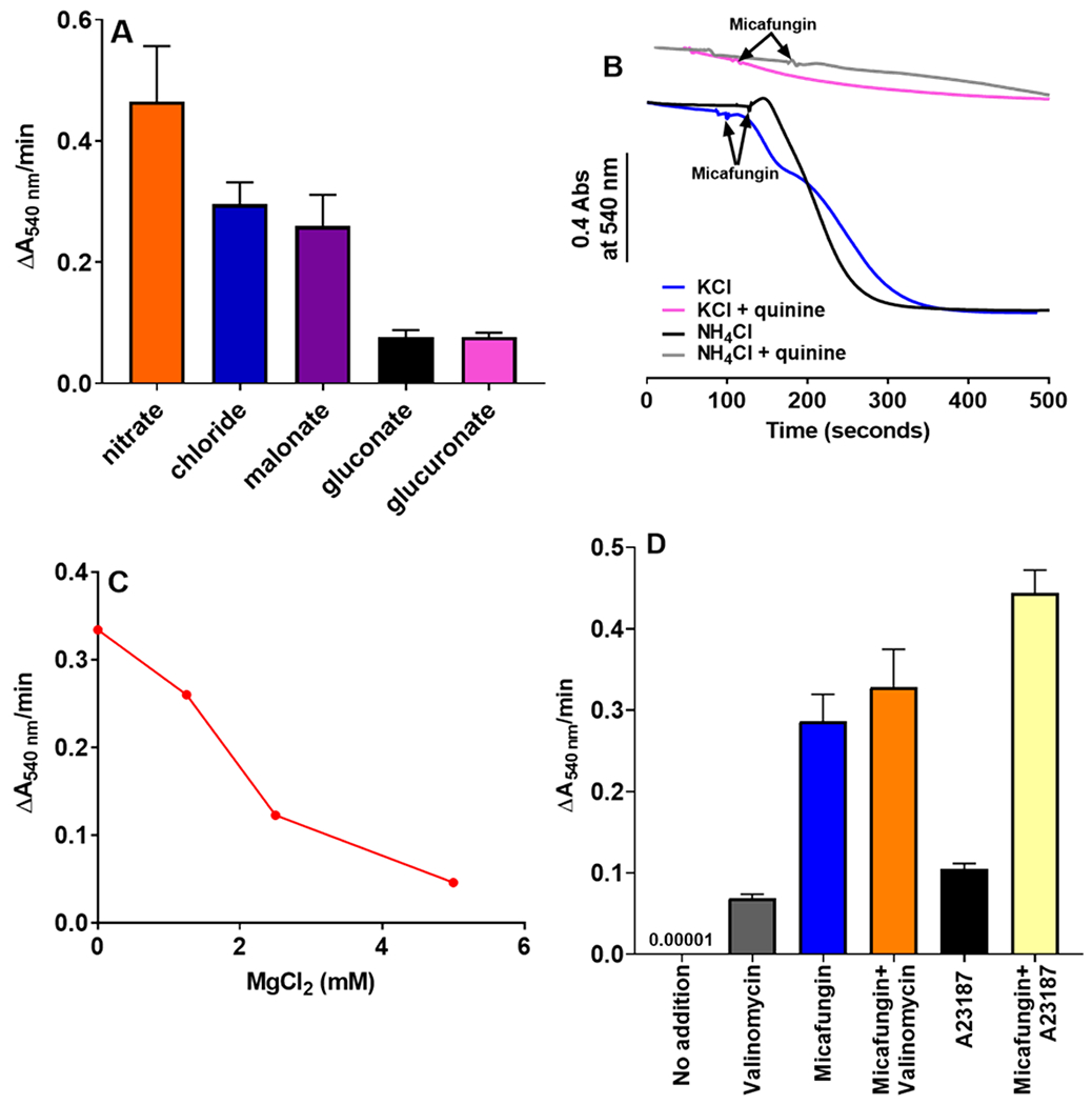
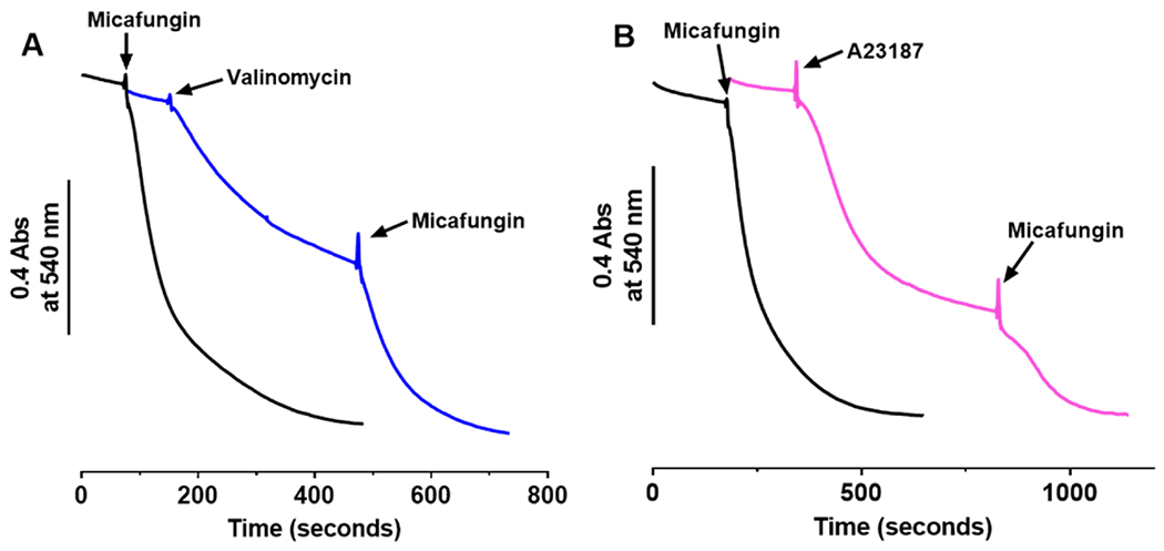
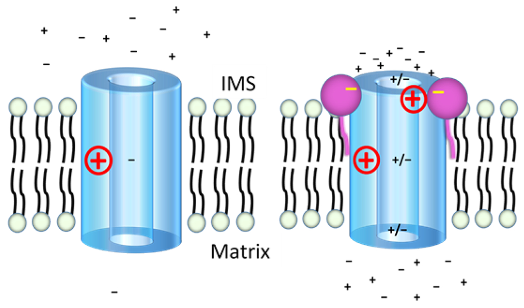
The antifungal activity of the drug micafungin, a cyclic lipopeptide that interacts with membrane proteins, may involve inhibition of fungal mitochondria. In humans, mitochondria are spared by the inability of micafungin to cross the cytoplasmic membrane. Using isolated mitochondria, we find that micafungin initiates the uptake of salts, causing rapid swelling and rupture of mitochondria with release of cytochrome c. The inner membrane anion channel (IMAC) is altered by micafungin to transfer both cations and anions. We propose that binding of anionic micafungin to IMAC attracts cations into the ion pore for the rapid transfer of ion pairs.

Keywords: Cyclic lipopeptide; Inner membrane anion channel; Ion channel; Micafungin; Mitochondrial ion channel; Mitochondrial respiratory chain complex; cytochrome c release.


Conflict of interest statement

Declaration of Competing Interest The authors declare that they have no known competing financial interests or personal relationships that could have appeared to influence the work reported in this paper.

Figures

Figure 1.
Figure 1.
Chemical structure of micafungin.
Figure 2.
Figure 2.
Micafungin causes the release of soluble cytochrome c from intact mitochondria. (A) Rates of O2 consumption by complex IV were measured at 25°C using an Oroboros FluoRespirometer in 2 mL of 50 mM MOPS, 5 mM KH2PO4, 100 mM KCl and 1 mM EGTA, pH 7.4. In the blue tracing of panel A, 0.7 mg of intact rat liver mitochondria were present at time zero. CIV activity was initiated by the reduction of endogenous cytochrome c with 3 mM ascorbate and 0.3 mM TMPD. The addition of 25 μM micafungin inhibited CIV activity, which was then restored by the addition of 7 μM horse heart cytochrome c. The pink tracing is identical to the blue tracing except that no micafungin was added. The addition of 7 μM horse cytochrome c has no effect indicating that the outer membrane of the mitochondria is intact. The black tracing was obtained using 0.7 mg of broken rat liver mitochondria (prepared as in Shirey et al (13)), where micafungin has access to both sides of the IMM. Ascorbate, TMPD and 25 μM horse heart cytochrome c were present from time zero and CIV O2 consumption was initiated by the addition of mitochondria. The addition of 25 μM micafungin stimulates CIV activity, as previously seen (13). (B) The assays of panel B were performed and labeled as explained for the blue and pink tracings of panel A, except that intact Candida mitochondria were used. (C) Reduced minus oxidized optical spectra were used to confirm that cytochrome c was released by treating intact rat liver (blue) or Candida (black) mitochondria with micafungin (see Methods). The α peak of reduced cytochrome c appears at 550 nm and the β peak at 520 nm.
Figure 3.
Figure 3.
(A) Measurements of mitochondrial light scattering show that 25 μM micafungin (blue trace) and 20 μg/ml alamethicin (black trace) induced large scale swelling of intact rat liver mitochondria (0.7 mg) suspended in 100 mM KCl, 5 mM KH2PO4, 1 mM EGTA and 50 mM MOPS, pH 7.4. (B) Evidence for the rupture of rat liver IMM during caused by MIMS is indicated by the release of the matrix enzyme citrate synthase, measured as described in Methods. The control and ‘+micafungin’ columns indicate the citrate synthase activity released after five minutes in the absence or presence of 25 μM micafungin, while the total CS column shows the entire citrate synthase activity present in an equivalent amount of intact mitochondria (n=3-4; error as SEM; ns=not significant).
Figure 4.
Figure 4.
(A) Micafungin (25 μM)-induced swelling of 0.6 mg rat liver mitochondria suspended in 100 mM KCl, 5 mM KH2PO4, 1mM EGTA, 50 mM MOPS, pH 7.4 at 25°C (blue tracing) can be halted by the addition of bovine serum albumin to 1 mg/ml (black tracing). (B) The initial rate of swelling (measured as in panel A, blue tracing) vs. the log of the concentration of micafungin shows that micafungin binding is cooperative (Hill slope of 2.9) with an EC50 of 22.5 μM.
Figure 5.
Figure 5.
Micafungin-induced mitochondrial swelling (MIMS) is distinct from the mitochondrial permeability transition pore (MPTP) since MIMS requires salt and MIMS is not inhibited by cyclosporine A. (A) When 0.4 mg intact, rat liver mitochondria are suspended in salt-free MSM buffer (220 mM mannitol, 70 mM sucrose and 10 mM MOPS pH 7.4) 0.5 μM tributyltin (TBT) induces rapid mitochondrial swelling (black tracing) by opening the MPTP (56). In MSM buffer without salt, 25 μM micafungin does not induce rapid swelling (blue tracing), but the CLP does induce swelling in MSM when 120 mM KCl is included (pink tracing). (B) Swelling induced by 25 μM micafungin in a buffer containing 0.3 mg rat liver mitochondria, 50 mM MOPS, 5 mM KH2PO4, 100 mM KCl and 1 mM EGTA, pH 7.4 (blue tracing) is not inhibited by the inclusion of 1 μM cyclosporine A prior to the addition of micafungin (black tracing). (A control for the inhibitory action of cyclosporine in our mitochondria is shown in Fig. S2.)
Figure 6.
Figure 6.
(A) The rate of MIMS vs the concentration of KCl. Intact rat liver mitochondria (0.4 mg) were suspended in a buffer of 5 mM KH2PO4, 1mM EGTA and 50 mM MOPS, pH 7.4, plus KCl as indicated. MIMS was initiated by the addition of 25 μM micafungin. (B) Neither the initial rate nor the extent of MIMS (measured as in panel A plus 100 mM KCl; 25 μM micafungin added where indicated) depends upon a membrane potential, as shown by nearly equal rates of swelling in the presence of 1 μM of the uncoupler FCCP (blue tracing; swelling rate of 0.237±0.031 ΔA540nm/min) and in the absence of FCCP (black tracing; swelling rate of 0.252±0.017 ΔA540nm/min).
Figure 7.
Figure 7.
(A) Comparing the initial rates of rat liver mitochondrial swelling induced by 25 μM micafungin in buffers containing 0.4 mg mitochondria, 5 mM KH2PO4, 1mM EGTA, 50 mM MOPS, pH 7.4, plus 100 mM of one of the following salts: potassium chloride, potassium gluconate, sodium nitrate, sodium malonate or sodium glucoronate. Error bars are SEM with an n=2-5. (B) Traces of micafungin-induced mitochondrial swelling, performed as in panel A, using either 100 mM KCl or 100 mM NH4Cl as the salt plus 500 μM quinine as indicated. (C) Swelling of rat liver mitochondria (0.5 mg) induced by 25 μM micafungin is inhibited by mM concentrations of MgCl2. (D) The initial rates of swelling by rat liver mitochondria (0.4 mg) in a buffer containing 55 mM KCl, 2.5 mM EDTA, 1 μM cyclosporine A, and 10 mM MOPS, pH 7.4. The additions indicated, used to initiate swelling, were 2 μM valinomycin, 25 μM micafungin and 4.75 μM A23187. Error bars are SEM with an n=2-5.
Figure 8.
Figure 8.
In buffer containing 0.4 mg mitochondria, 55 mM KCl, 1 μM cyclosporine A, 2.5 mM EDTA, and 10 mM MOPS, pH 7.4, mitochondrial swelling in the presence of 2 μM valinomycin (A) or 4.75 μM A23187 (B, pink) begins to plateau before complete mitochondrial rupture but swelling resumes upon the addition of 25 μM micafungin. Traces of mitochondrial swelling induced by only 25 μM micafungin are shown in black.
Figure 9.
Figure 9.
A proposed model for how micafungin stimulates both cation and anion transfer through inner membrane anion channel (IMAC) of mitochondria. While the structure of IMAC remains unknown, IMAC is depicted as a cylinder with a wide pore because the channel transfers ions with a wide range of sizes. A positive charge halfway through the pore indicates an electropositive filter (such as a lysine side chain) that many anion channels employ to limit cation transfer. Since MIMS does not respond to the presence or absence of an initial membrane potential (see text), no potential is indicated. The left panel shows IMAC in the absence of micafungin as an anion channel of low conductance. The right panel posits that the addition of micafungin allows several micafungin molecules (only two are shown) to bind around the IMS entry of the pore of IMAC. The negative charges of the bound micafungin molecules attract mobile cations to the pore entry, the mobile cations attract mobile anions and a resulting increase in the local concentration of ions allows formation of cation/anion pairs that avoid rejection by electropositive filters as they transit the IMAC pore. The micafungin molecule on the right is shown to be neutralizing a positive side chain near the IMS mouth of the pore, as may be the case if a CLIC5 protein is IMAC (see text). This would also allow an increase in the concentration of cations near the pore entry. The net direction (into the matrix) and the energy for ion transfer is provided by the chemical potential for anions.

Similar articles

References

    1. Akar FG, Aon MA, Tomaselli GF, O’Rourke B, 2005. The mitochondrial origin of postischemic arrhythmias. J Clin Invest 115, 3527–3535, https://www.ncbi.nlm.nih.gov/pubmed/16284648. - PMC - PubMed
    1. Aon MA, Cortassa S, Maack C, O’Rourke B, 2007. Sequential opening of mitochondrial ion channels as a function of glutathione redox thiol status. J Biol Chem 282, 21889–21900, https://www.ncbi.nlm.nih.gov/pubmed/17540766. - PMC - PubMed
    1. Aon MA, Cortassa S, Marban E, O’Rourke B, 2003. Synchronized whole cell oscillations in mitochondrial metabolism triggered by a local release of reactive oxygen species in cardiac myocytes. J Biol Chem 278, 44735–44744, https://www.ncbi.nlm.nih.gov/pubmed/12930841. - PubMed
    1. Balleza D, Alessandrini A, Beltran Garcia MJ, 2019. Role of Lipid Composition, Physicochemical Interactions, and Membrane Mechanics in the Molecular Actions of Microbial Cyclic Lipopeptides. J Membr Biol 252, 131–157, https://www.ncbi.nlm.nih.gov/pubmed/31098678. - PubMed
    1. Beavis AD, 1992. Properties of the inner membrane anion channel in intact mitochondria. J Bioenerg Biomembr 24, 77–90, https://www.ncbi.nlm.nih.gov/pubmed/1380509. - PubMed

Publication types

LinkOut - more resources