. 2023 Jul 15:332:199119.
doi: 10.1016/j.virusres.2023.199119. Epub 2023 May 26.

PQBP1 regulates the cellular inflammation induced by avian reovirus and interacts with the viral p17 protein

Affiliations

PQBP1 regulates the cellular inflammation induced by avian reovirus and interacts with the viral p17 protein

Chengcheng Zhang et al. Virus Res. .

Abstract

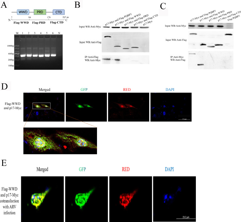
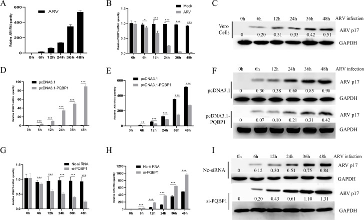
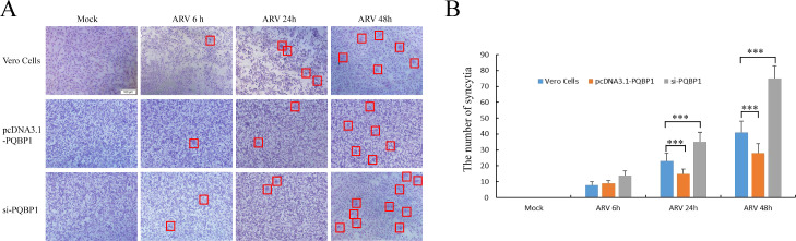
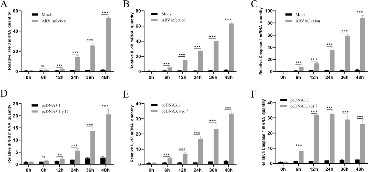
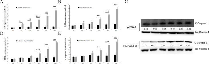
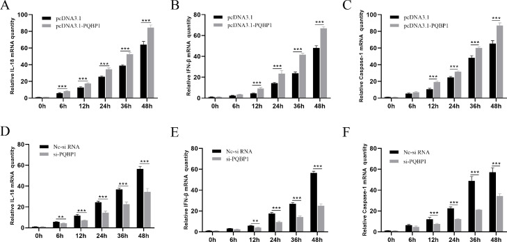
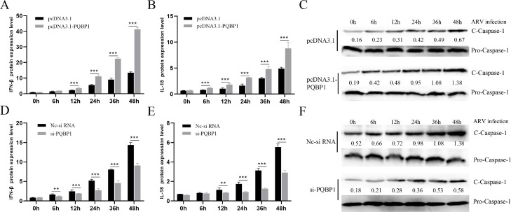
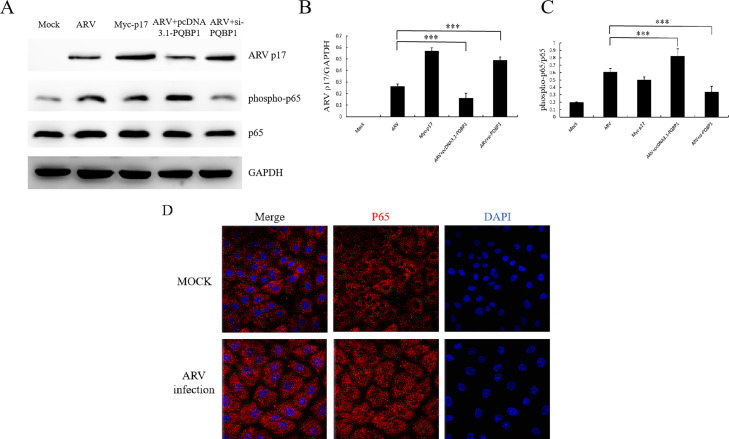
Avian reovirus (ARV) can commonly infect a flock and cause immunosuppressive diseases in poultry. The nonstructural protein p17 is involved in viral replication, and significant progress has been made in showing its ability to regulate cellular signaling pathways. In our previous study, to further investigate the effect of ARV p17 protein on viral replication, the host protein polyglu-tamine binding protein 1 (PQBP1) was identified to interact with p17 by a yeast two-hybrid system. In the current study, the interaction between PQBP1 and p17 protein was further confirmed by laser confocal microscopy and coimmunoprecipitation assays. In addition, the N-terminal WWD of PQBP1 was found to mediate the process of binding to the p17 protein. Interestingly, we found that ARV infection significantly inhibited PQBP1 expression. While the quantity of ARV replication was largely influenced by PQBP1, PQBP1 overexpression decreased ARV replication. In contrast, upon PQBP1 knockdown, the quantity of ARV was notably increased. ARV infection and p17 protein expression were both proven to induce PQBP1 to mediate cellular inflammation. In the current study, we revealed through qRT‒PCR, ELISA and Western blotting methods that PQBP1 plays a positive role in ARV-induced inflammation. Furthermore, the mechanism of this process was shown to involve the NFκB-dependent transcription of inflammatory genes. In addition, PQBP1 was shown to regulate the phosphorylation of p65 protein. In conclusion, this research provides clues to elucidating the function of the p17 protein and the pathogenic mechanism of ARV, especially the cause of the inflammatory response. It also provides new ideas for the study of therapeutic targets of ARV.

Keywords: Avian reovirus; Inflammation; P17 protein; PQBP1.


Conflict of interest statement

Declaration of Competing Interest The authors declare that they have no conflict of interest!

Figures

Fig. 1
Fig. 1
The N-terminus of PQBP1 interacts with the ARV p17 protein. (A) Schematic representation of the protein domains (shown as amino acids number) of PQBP1 and the PCR results of each domain. M, DL1000 marker; 1–2, Flag-WWD(313 bp); 3–4, Flag-PRD(298 bp); 5–6, Flag-CTD(313 bp); N, Negative control. (B) The N-terminal region of PQBP1 interacts with the p17 protein. 293T cells were transfected with different portions or the full-length form of PQBP1 with a Flag tag and p17-Myc plasmids for 48 h and harvested. Cell lysates were immunoprecipitated with an antibody against Flag followed by Western blotting analysis. (C) Reciprocal co-IP experiments showed that the anti-Myc antibody precipitated Flag-WWD. (D) Colocalization of WWD of PQBP1 and ARV p17 protein. 293T cells were cotransfected with Flag-WWD and p17-Myc for 24 h, then incubated with anti-Flag and anti-Myc primary antibodies and immunostained with PE- or FITC-labeled secondary antibodies. Finally, the cells were analyzed by confocal microscopy. (E) Colocalization of Flag-WWD and p17-Myc in the condition of ARV infection.
Fig. 2
Fig. 2
Cellular PQBP1 inhibits ARV proliferation. (A) Vero cells were infected with ARV at an MOI of 1 for different times, and the quantity of ARV replication was assayed by qRT‒PCR. (B) Intracellular PQBP1 expession after ARV infection was revealed by qRT‒PCR. (C) The ARV p17 protein levels in Vero cells were analyzed by Western blotting. The numbers represent the ratio of p17/GAPDH in different ARV infection time. (D) Intracellular PQBP1 expression after transfection with the pcDNA3.1-PQBP1 plasmid in Vero cells was revealed by qRT‒PCR. (E) After transfected with pcDNA3.1-PQBP1 48 h, the Vero cells infeciton ARV at indicated time. The quantity of ARV replication in Vero cells was revealed by qRT‒PCR. (F) The ARV p17 protein level in PQBP1-overexpressing Vero cells was analyzed by Western blotting. The numbers represent the ratio of p17/GAPDH in different ARV infection time. (G) Intracellular PQBP1 expression after transfection with si-PQBP1 or Nc-si RNA, then infection with ARV with MOI of 1 at different time in Vero cells was revealed by qRT‒PCR. (H) After transfected with si-PQBP1 48 h, the Vero cells infeciton ARV at indicated time. The quantity of ARV replication in Vero cells was revealed by qRT‒PCR. (I) The ARV p17 protein level in PQBP1-knockdown Vero cells was analyzed by Western blotting. The numbers represent the ratio of p17/GAPDH in different ARV infection time. Results are presented as the means ± SD of data from three independent experiments. (*P < 0.05, **P < 0.01; and ***P < 0.001).
Fig. 3
Fig. 3
Cellular PQBP1 inhibits ARV proliferation in cell level. (A) The level of syncytium formation in Vero cells was detected by Giemsa-staining at different ARV infection times. Images were captured on an inverted microscope at 10 × objective. (B) The number of syncytia per microscopic field in Vero cells. Results are presented as the means ± SD of data from three independent experiments. ***, P < 0.001.
Fig. 4
Fig. 4
ARV infection and expression of p17 protein both induce cellular inflammation. Vero cells were infected with ARV at an MOI of 1 or expressing ARV p17 protein for different times, and the inflammatory factors were assayed by qRT-PCR. (A), (B) and (C) show the effect of ARV infection on the mRNA transcription levels of IFN-β, IL-18 and caspase-1, respectively. (D), (E) and (F) show the effect of ARV p17 protein expression on the mRNA transcription levels of IFN-β, IL-18 and caspase-1, respectively. Statistical analysis: unpaired t-test (*P < 0.05, **P < 0.01; and ***P < 0.001).
Fig. 5
Fig. 5
ARV infection and expression of p17 protein both induce cellular inflammation. Vero cells were infected with ARV at an MOI of 1 or expressing ARV p17 protein for different times, and the inflammatory factors were assayed by ELISA or Western blotting. (A) and (B) show the effect of ARV infection on the protein levels (ng/ul) of IFN-β and IL-18, respectively. (D), (E) and (C) show the effect of p17 protein expression on the protein levels (ng/ul) of IFN-β, IL-18 and Caspase-1, respectively. Results are presented as the means ± SD of data from three independent experiments. (***P < 0.001).
Fig. 6
Fig. 6
PQBP1 plays a positive role in ARV-induced inflammation. Vero cells with PQBP1 overexpression or knockdown were then infected with ARV at an MOI of 1 for different times, and the inflammatory factors were assayed by qRT-PCR. (A), (B) and (C) show the effect of PQBP1 overexpression on the mRNA transcription levels of IL-18, IFN-β and caspase-1, respectively. (D), (E) and (F) show the effect of PQBP1 knockdown on the mRNA transcription levels of IFN-β, IL-18 and caspase-1, respectively. Results are presented as the means ± SD of data from three independent experiments. (*P < 0.05, **P < 0.01; and ***P < 0.001).
Fig. 7
Fig. 7
PQBP1 plays a positive role in ARV-induced inflammation. Vero cells with PQBP1 overexpression or knockdown were then infected with ARV at an MOI of 1 for different times, and the inflammatory factors were assayed by ELISA or Western blotting. (A), (B) and (C) show the effect of PQBP1 overexpression on the protein levels (ng/ul) of IFN-β, IL-18 and Caspase-1, respectively. (D), (E) and (F) show the effect of PQBP1 knockdown on the protein levels (ng/ul) of IFN-β, IL-18 and Caspase-1, respectively. Results are presented as the means ± SD of data from three independent experiments. (*P < 0.05, **P < 0.01; and ***P < 0.001).
Fig. 8
Fig. 8
PQBP1 mediates ARV-induced NF-κB activation in Vero cells. (A) Western blots reveal activation of NFκB (phospho-p65) after ARV infection, p17 protein expression or overexpression or PQBP1 knockdown in Vero cells. (B) Relative p17 protein levels are presented. (C) Relative phospho-p65 protein levels are presented. This experiment was repeated independently three times with similar results. (D) Immunofluorescence images of Vero cells stained for p65 after ARV infection 24 h or mock infection. Scale bars: 49.1 µm. Results are presented as the means ± SD of data from three independent experiments. (***P < 0.001).

Similar articles

Cited by

References

    1. Benavente J., Martinez-Costas J. Avian reovirus: structure and biology. Virus Res. 2007;123(2):105–119. - PubMed
    1. Choi Y.R., Kim S.W., Shang K., Park J.Y., Zhang J.F., Jang H.K., Wei B., Cha S.Y., Kang M. Avian reoviruses from wild birds exhibit pathogenicity to specific pathogen free chickens by footpad route. Front. Vet. Sci. 2022;9 - PMC - PubMed
    1. Clarke P., Debiasi R.L., Goody R., Hoyt C.C., Richardson-Burns S., Tyler K.L. Mechanisms of reovirus-induced cell death and tissue injury: role of apoptosis and virus-induced perturbation of host-cell signaling and transcription factor activation. Viral Immunol. 2005;18(1):89–115. - PMC - PubMed
    1. Drummond T.G., Hill M.G., Barton J.K. Electrochemical DNA sensors. Nat. Biotechnol. 2003;21(10):1192–1199. - PubMed
    1. He T.S., Dang L., Zhang J., Zhang J., Wang G., Wang E., Xia H., Zhou W., Wu S., Liu X. The Hippo signaling component LATS2 enhances innate immunity to inhibit HIV-1 infection through PQBP1-cGAS pathway. Cell Death Differ. 2022;29(1):192–205. - PMC - PubMed

Publication types