Clinical Validation of Stimulated Raman Histology for Rapid Intraoperative Diagnosis of Central Nervous System Tumors
- PMID: 37201685
- PMCID: PMC10527246
- DOI: 10.1016/j.modpat.2023.100219
Clinical Validation of Stimulated Raman Histology for Rapid Intraoperative Diagnosis of Central Nervous System Tumors
Abstract
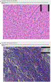
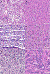
Stimulated Raman histology (SRH) is an ex vivo optical imaging method that enables microscopic examination of fresh tissue intraoperatively. The conventional intraoperative method uses frozen section analysis, which is labor and time intensive, introduces artifacts that limit diagnostic accuracy, and consumes tissue. SRH imaging allows rapid microscopic imaging of fresh tissue, avoids tissue loss, and enables remote telepathology review. This improves access to expert neuropathology consultation in both low- and high-resource practices. We clinically validated SRH by performing a blinded, retrospective two-arm telepathology study to clinically validate SRH for telepathology at our institution. Using surgical specimens from 47 subjects, we generated a data set composed of 47 SRH images and 47 matched whole slide images (WSIs) of formalin-fixed, paraffin-embedded tissue stained with hematoxylin and eosin, with associated intraoperative clinicoradiologic information and structured diagnostic questions. We compared diagnostic concordance between WSI and SRH-rendered diagnoses. Also, we compared the 1-year median turnaround time (TAT) of intraoperative conventional neuropathology frozen sections with prospectively rendered SRH-telepathology TAT. All SRH images were of sufficient quality for diagnostic review. A review of SRH images showed high accuracy in distinguishing glial from nonglial tumors (96.5% SRH vs 98% WSIs) and predicting final diagnosis (85.9% SRH vs 93.1% WSIs). SRH-based diagnosis and WSI-permanent section diagnosis had high concordance (κ = 0.76). The median TAT for prospectively SRH-rendered diagnosis was 3.7 minutes, approximately 10-fold shorter than the median frozen section TAT (31 minutes). The SRH-imaging procedure did not affect ancillary studies. SRH generates diagnostic virtual histologic images with accuracy comparable to conventional hematoxylin and eosin-based methods in a rapid manner. Our study represents the largest and most rigorous clinical validation of SRH to date. It supports the feasibility of implementing SRH as a rapid method for intraoperative diagnosis complementary to conventional pathology laboratory methods.
Keywords: artificial intelligence; brain tumors; frozen section; glioma; intraoperative diagnosis; stimulated Raman histology.
Copyright © 2023 United States & Canadian Academy of Pathology. Published by Elsevier Inc. All rights reserved.
Figures


References
Publication types
MeSH terms
Substances
Grants and funding
LinkOut - more resources
Full Text Sources

