. 2023 Aug;16(3):225-235.
doi: 10.21053/ceo.2023.00227. Epub 2023 May 18.

Effects of Particulate Matter Exposure on the Eustachian Tube and Middle Ear Mucosa of Rats

Affiliations

Effects of Particulate Matter Exposure on the Eustachian Tube and Middle Ear Mucosa of Rats

Hyun Min Lee et al. Clin Exp Otorhinolaryngol. 2023 Aug.

Abstract

Objectives: Particulate matter (PM) is a risk factor for various diseases. Recent studies have established an association between otitis media (OM) and PM exposure. To confirm this relationship, we developed a novel exposure model designed to control the concentration of PM, and we observed the effects of PM exposure on the Eustachian tube (ET) and middle ear mucosa of rats.

Methods: Forty healthy, 10-week-old, male Sprague-Dawley rats were divided into 3-day, 7-day, 14-day exposure, and control groups (each, n=10). The rats were exposed to incense smoke as the PM source for 3 hours per day. After exposure, bilateral ETs and mastoid bullae were harvested, and histopathological findings were compared using microscopy and transmission electron microscopy (TEM). The expression levels of interleukin (IL)-1β, IL-6, tumor necrosis factor-α, and vascular endothelial growth factor (VEGF) in the middle ear mucosa of each group were compared using real-time reverse transcription polymerase chain reaction (RT-PCR).

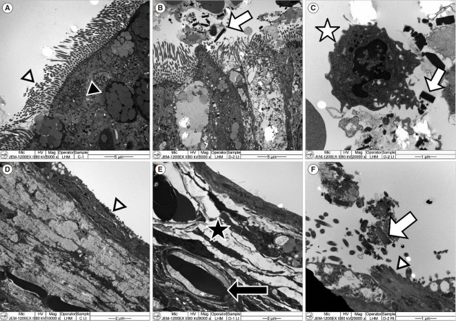
Results: In the ET mucosa of the exposure group, the goblet cell count significantly increased after PM exposure (P=0.032). In the middle ear mucosa, subepithelial space thickening, increased angio-capillary tissue, and inflammatory cell infiltration were observed. Moreover, the thickness of the middle ear mucosa in the exposure groups increased compared to the control group (P<0.01). The TEM findings showed PM particles on the surface of the ET and middle ear mucosa, and RT-PCR revealed that messenger RNA (mRNA) expression of IL-1β significantly increased in the 3-day and 7-day exposure groups compared to the control group (P=0.035). VEGF expression significantly increased in the 7-day exposure group compared to the control and 3-day exposure groups (P<0.01).

Conclusion: The ET and middle ear mucosa of rats showed histopathologic changes after acute exposure to PM that directly reached the ET and middle ear mucosa. Therefore, acute exposure to PM may play a role in the development of OM.

Keywords: Eustachian Tube; Incense; Middle Ear; Otitis Media; Particulate Matter; Rats.


Conflict of interest statement

No potential conflict of interest relevant to this article was reported.

Figures

Fig. 1.
Fig. 1.
Schematic overview of the research process. TEM, transmission electron microscopy; RT-PCR, real-time reverse transcription polymerase chain reaction; IL, interleukin.
Fig. 2.
Fig. 2.
Particulate matter exposure model for rats. The blower supplies room air to the incense burning and rat exposure chambers. The incense smoke from the incense burning chamber is supplied to the rat exposure chamber, and the internal environment is monitored using the particulate matter (PM) monitor and temperature and humidity monitor. The amount of PM is controlled by the airflow regulator. Air suction is installed in the rat exposure chamber for continuous ventilation.
Fig. 3.
Fig. 3.
Measurement of the middle ear mucosa thickness in the control group under light microscopy (H&E, ×12.5). The average thickness of the middle ear mucosa was calculated by measuring the thickness at three sites (black circles): one at the mucosa on the contralateral side of the cochlea (black triangle), and the other two at the bilateral mucosa perpendicular to the imaginary line between the cochlea and the contralateral mucosa.
Fig. 4.
Fig. 4.
Histopathologic features of the Eustachian tube (ET) mucosa. (A) Light microscope findings of the ET in the control group (H&E, ×100). The pseudostratified mucociliary respiratory-like epithelium is observed (arrowheads). (B) High magnification image of ET mucosa in the control group (H&E, ×400). Cilia of the epithelial cell (arrow) and goblet cells (arrowheads) are shown on the ET mucosa. (C) Goblet cells (arrowheads) in the ET mucosa are observed in the control group (Alcian blue stain, ×100). (D) Light microscope findings of the ET in the 3-day exposure group (H&E, ×100). (E) High magnification image of ET mucosa in the 3-day exposure group (H&E, ×400). An increased number of hypertrophied goblet cells (arrowheads) is shown. (F) An increased number of goblet cells (arrowheads) of ET mucosa is observed in the 3-day exposure group (Alcian blue stain, ×100).
Fig. 5.
Fig. 5.
Histopathologic findings (H&E) of the middle ear mucosa. In the control group: (A, ×100): Non-keratinized stratified squamous epithelium (arrowheads). (B, ×400): The epithelial layer (arrowhead) shows a dense configuration. In the 3-day exposure group: (C, ×100): Hyperplasia of the stratified squamous cells (arrow) and increased cilia (arrowhead). (D, ×400): Hyperplasia of epithelial cells, thickening of the subepithelial space, and enlargement of angio-capillary tissue (arrow). In the 7-day exposure group: (E, ×100): Increased angio-capillary tissue within the thickened subepithelial tissue (arrow). (F, ×400): Inflammatory cell (lymphocyte) infiltration (arrow). In the 14-day exposure group: (G, ×100): A thickened subepithelial space and increased angio-capillary tissue (arrow). (H, ×400): Increased cilia (arrowhead) and inflammatory cell (lymphocyte) infiltration (arrow).
Fig. 6.
Fig. 6.
Alterations in the thickness of the middle ear mucosa. Compared to the thickness of the middle ear mucosa in the control group, that in the 3-day, 7-day, and 14-day exposure groups significantly increased (P=0.009, P=0.002, and P=0.001, respectively). The duration of particulate matter exposure and the thickness of the mucous membrane did not show a proportional trend. × indicates mean value; horizontal line in the box, median value. *P<0.05.
Fig. 7.
Fig. 7.
Transmission electron microscopy findings of the Eustachian tube (ET) and middle ear mucosa. (A) In the ET mucosa of the control group, cilia (white arrowhead) and goblet cells (black arrowhead) were observed (×5,000). (B) In the ET mucosa of the 3-day exposure group, particulate matter (PM) particles (arrow) were observed over the cilia (×5,000). (C) In the ET mucosa of the 3-day exposure group with higher magnification, a neutrophil (asterisk) and PM particles (arrow) were observed together (×20,000). (D) In the middle ear mucosa of the control group, small ciliary tissue (arrowhead) was observed (×10,000). (E) In the middle ear mucosa of the 3-day exposure group, thickened subepithelial tissue (asterisk) was identified, and erythrocytes (arrow) were observed inside of the angio-capillary tissue (×8,000). (F) In the middle ear mucosa near the ET in the 3-day exposure group, cilia (arrowhead) and PM particles (arrow) over the cilia were observed (×20,000).
Fig. 8.
Fig. 8.
Cytokine expression by the control, 3-day, 7-day, and 14-day exposure groups by real-time polymerase chain reaction. The messenger RNA (mRNA) expression of interleukin (IL)-1β (A) significantly increased after particulate matter (PM) exposure in the 3-day and 7-day exposure groups (P=0.035 by the Kruskal-Wallis test). The mRNA expression of IL-6 (B) and tumor necrosis factor (TNF)-α (C) did not significantly increase after PM exposure in the 3-day, 7-day, and 14-day exposure groups (P=0.142 and P=0.472, respectively, by the Kruskal-Wallis test). The mRNA expression of vascular endothelial growth factor (VEGF; D) significantly increased after PM exposure in the 7-day exposure group (P<0.01 by the Kruskal-Wallis test). × indicates mean value; horizontal line in the box, median value. *P<0.05.

References

    1. World Health Organization . World Health Organization; 2022. Ambient (outdoor) air pollution [Internet] [cited 2023 May 1]. Available from: https://www.who.int/news-room/fact-sheets/detail/ambient-(outdoor)-air-q....
    1. Thompson JE. Airborne particulate matter: human exposure and health effects. J Occup Environ Med. 2018 May;60(5):392–423. - PubMed
    1. Park M, Han J, Jang MJ, Suh MW, Lee JH, Oh SH, et al. Air pollution influences the incidence of otitis media in children: a national population-based study. PLoS One. 2018 Jun;13(6):e0199296. - PMC - PubMed
    1. Naclerio R, Ansotegui IJ, Bousquet J, Canonica GW, D’Amato G, Rosario N, et al. International expert consensus on the management of allergic rhinitis (AR) aggravated by air pollutants: impact of air pollution on patients with AR: current knowledge and future strategies. World Allergy Organ J. 2020 Apr;13(3):100106. - PMC - PubMed
    1. Park M, Han J, Park J, Jang MJ, Park MK. Particular matter influences the incidence of acute otitis media in children. Sci Rep. 2021 Oct;11(1):19730. - PMC - PubMed