. 2023 Jun 28;194(1):109-119.
doi: 10.1093/toxsci/kfad048.

Role of PPARγ in dyslipidemia and altered pulmonary functioning in mice following ozone exposure

Affiliations

Role of PPARγ in dyslipidemia and altered pulmonary functioning in mice following ozone exposure

Ley Cody Smith et al. Toxicol Sci. .

Abstract

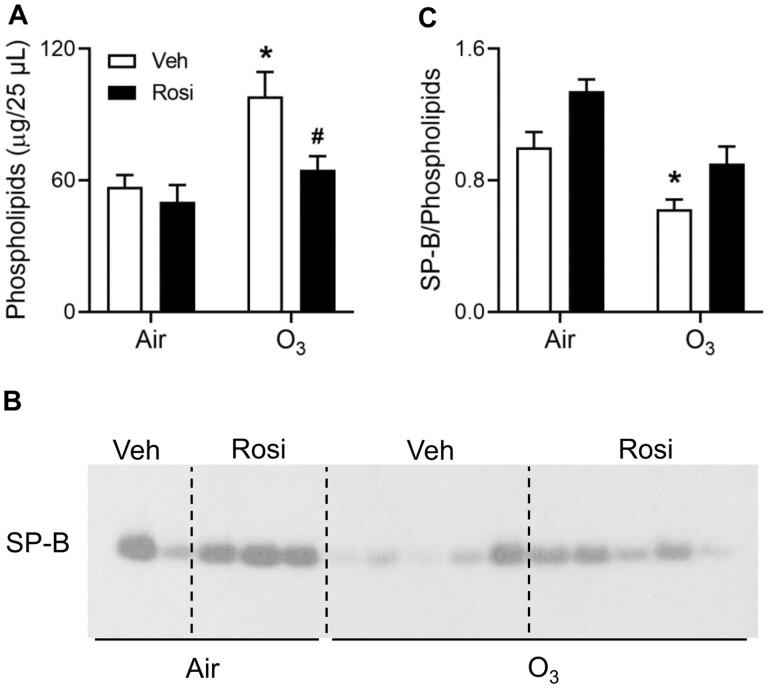
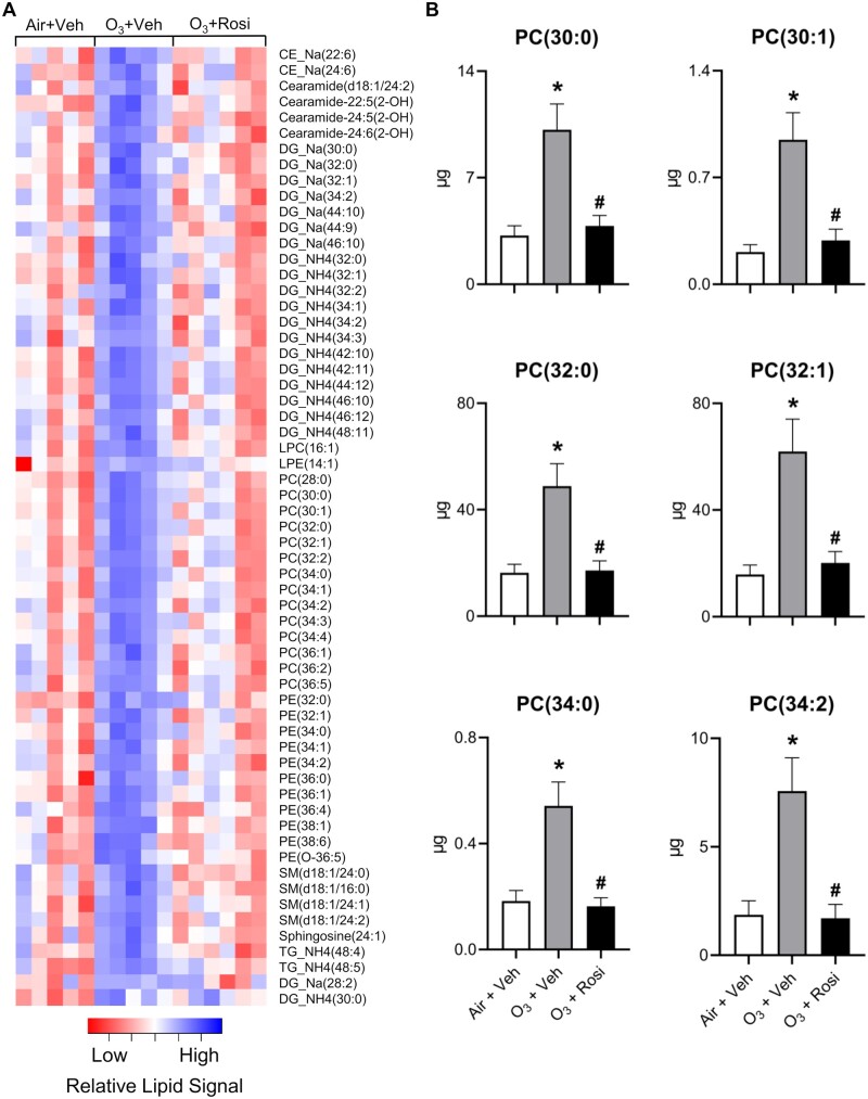
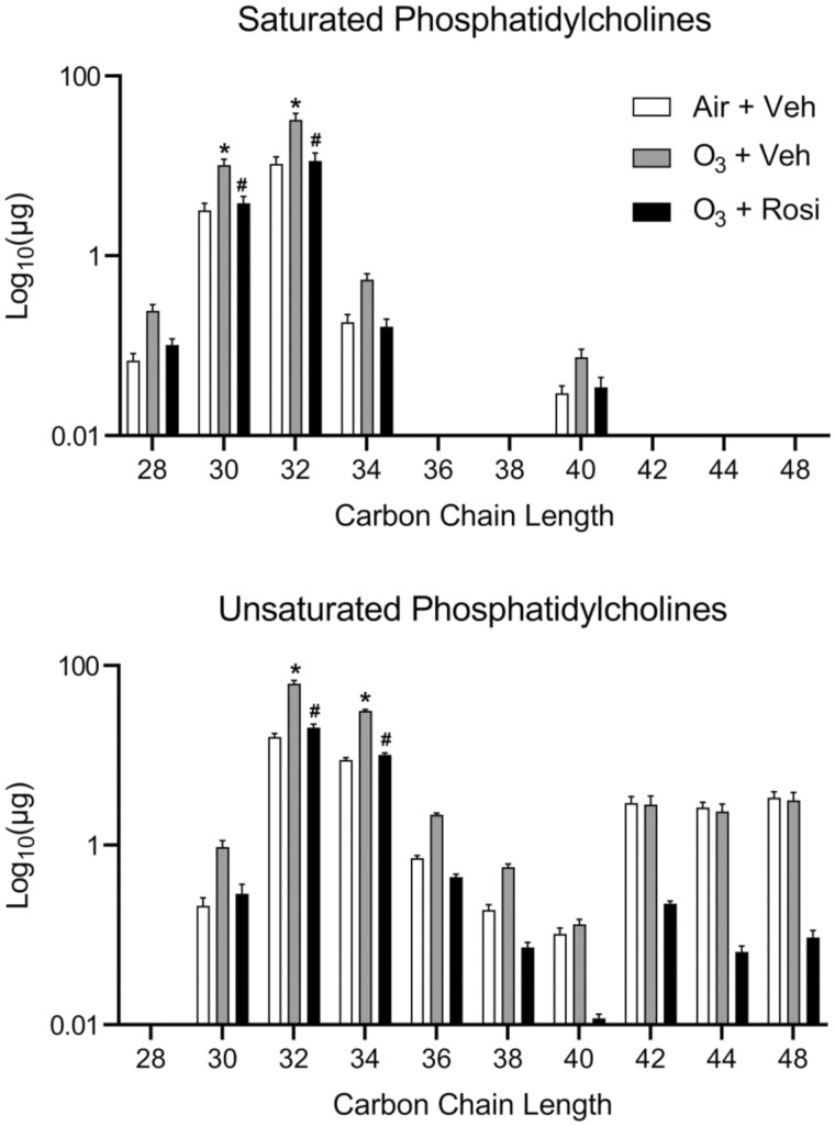
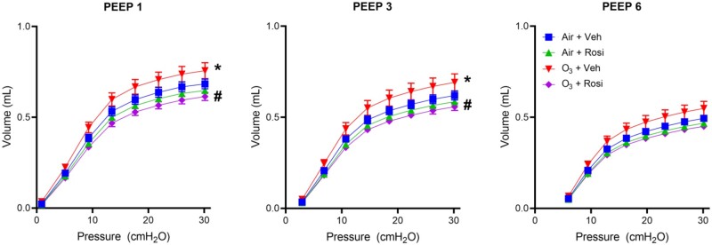
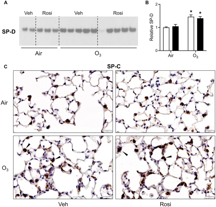
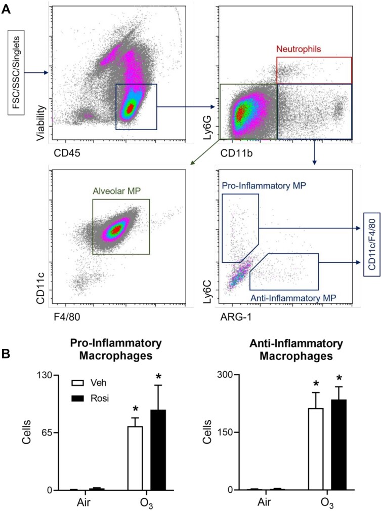
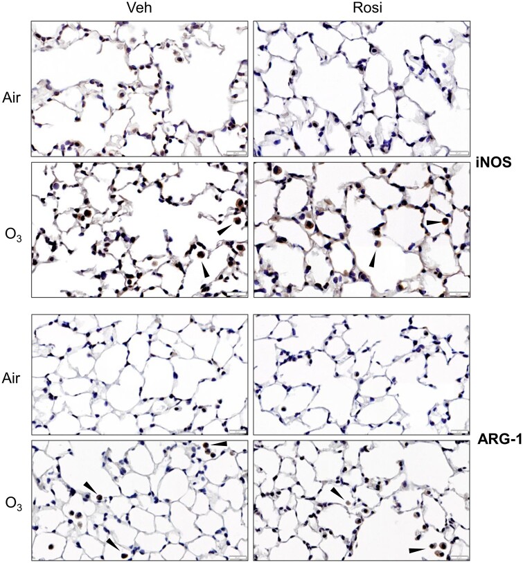
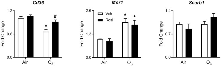
Exposure to ozone causes decrements in pulmonary function, a response associated with alterations in lung lipids. Pulmonary lipid homeostasis is dependent on the activity of peroxisome proliferator activated receptor gamma (PPARγ), a nuclear receptor that regulates lipid uptake and catabolism by alveolar macrophages (AMs). Herein, we assessed the role of PPARγ in ozone-induced dyslipidemia and aberrant lung function in mice. Exposure of mice to ozone (0.8 ppm, 3 h) resulted in a significant reduction in lung hysteresivity at 72 h post exposure; this correlated with increases in levels of total phospholipids, specifically cholesteryl esters, ceramides, phosphatidylcholines, phosphorylethanolamines, sphingomyelins, and di- and triacylglycerols in lung lining fluid. This was accompanied by a reduction in relative surfactant protein-B (SP-B) content, consistent with surfactant dysfunction. Administration of the PPARγ agonist, rosiglitazone (5 mg/kg/day, i.p.) reduced total lung lipids, increased relative amounts of SP-B, and normalized pulmonary function in ozone-exposed mice. This was associated with increases in lung macrophage expression of CD36, a scavenger receptor important in lipid uptake and a transcriptional target of PPARγ. These findings highlight the role of alveolar lipids as regulators of surfactant activity and pulmonary function following ozone exposure and suggest that targeting lipid uptake by lung macrophages may be an efficacious approach for treating altered respiratory mechanics.

Keywords: PPARγ; macrophage; ozone; pulmonary lipids.


Figures

Figure 1.
Figure 1.
Effects of rosiglitazone (Rosi) on ozone (O3)-induced alterations in lung phospholipids and SP-B. Lung lining fluid was collected 72 h after exposure of mice to Air + Vehicle (Veh), Air + Rosi, O3 + Veh, or O3 + Rosi. A, Total phospholipids were analyzed in lung lining fluid. B, Equal amounts of phospholipids were loaded into each well of a denaturing gel. SP-B levels were assessed by western blotting. Each lane represents one mouse; data shown are representative of 3 independent experiments. C, Western blots were quantified using ImageJ. Band intensities were normalized to the Air + Veh group within individual blots and then averaged across the blots; data are expressed relative to Air + Veh. Bars, mean ± SE (n = 7–16 mice). Data were analyzed by 2-way ANOVA followed by Tukey’s multiple comparisons test. *Significantly different (p < .05) from Air + Veh. #Significantly different (p < .05) from O3 + Veh.
Figure 2.
Figure 2.
Rosi suppresses O3-induced alterations in lung lipids. Lipidomic profiles in lung lining fluid were assessed 72 h after exposure of mice to Air + Vehicle (Veh), O3 + Veh, or O3 + Rosi by non-targeted mass spectrometry as described in the Materials and methods section. A, Heatmap of lipid species that exhibited significant differences in ion intensities between Air + Veh and O3 + Veh groups, as determined by Student’s t test (p < .05). Colors indicate mean-centered Log10(Ion Intensities). B, Absolute quantitation of phosphatidylcholine species including 1,2-dipentadecanoyl-sn-glycero-3-phosphocholine [PC(30:0)], 1-dodecanoyl-2-(9Z-octadecenoyl)-sn-glycero-3-phosphocholine [PC(30:1)], 1,2-dihexadecanoyl-sn-glycero-3-phosphocholine [PC(32:0)], 1-tetradecanoyl-2-(11Z-octadecenoyl)-sn-glycero-3-phosphocholine [PC(32:1)], 1-decanoyl-2-tetracosanoyl-sn-glycero-3-phosphocholine [PC(34:0)], 1-hexadecanoyl-2-(10E, 12Z-octadecadienoyl)-sn-glycero-3-phosphocholine [PC(34:2)] based on deuterated-PC(32:0) spike-in. Bars, mean ± SE (n = 5–6). Data were analyzed by 1-way ANOVA followed by Tukey’s multiple comparisons test. *Significantly different (p < .05) from Air + Veh. #Significantly different (p < .05) from O3 + Veh.
Figure 3.
Figure 3.
Effects of Rosi on O3-induced alterations in phosphatidylcholine species. Total amounts of saturated and unsaturated phosphocholine species were assessed in lung lining fluid 72 h after exposure of mice to Air + Vehicle (Veh), O3 + Veh, or O3 + Rosi by non-targeted mass spectrometry as described in the Materials and methods section. Absolute phosphatidylcholine levels were calculated based on deuterated-PC(32:0) spike-in. Bars, mean ± SE (n = 5–6). Data were analyzed by 2-way ANOVA followed by Tukey’s multiple comparisons test. *Significantly different (p < .05) from Air + Veh. #Significantly different (p < .05) from O3 + Veh.
Figure 4.
Figure 4.
Effects of Rosi on O3-induced alterations in respiratory mechanics. Pulmonary function was analyzed using a SCIREQ flexiVent system 72 h after exposure of mice to Air + Vehicle (Veh), Air + Rosi, O3 + Veh, or O3 + Rosi. PV loops were generated at PEEPs of 1, 3, and 6 cm H2O. Data are mean ± SE (n = 4). Data were analyzed by 2-way ANOVA. *Significantly different (p < .05) from Air + Veh. #Significantly different (p < .05) from O3 + Veh.
Figure 5.
Figure 5.
Effects of Rosi on O3-induced alterations in SP-D and pro-SP-C. A, SP-D levels were assessed 72 h after exposure of mice to Air + Vehicle (Veh), Air + Rosi, O3 + Veh, or O3 + Rosi by western blotting. A, Representative gel from 3 independent experiments is shown. Each lane represents one mouse. B, Blots were quantified using ImageJ. Band intensities were normalized to the Air + Veh group within individual blots and then averaged across the blots; data are expressed relative to Air + Veh. Bars, mean ± SE (n = 7–10 mice). Data were analyzed by 2-way ANOVA followed by Tukey’s multiple comparisons test. *Significantly different (p < .05) from Air + Veh. C, Tissue sections collected 72 h after exposure were stained with antibody to pro-SP-C. Binding was visualized using a peroxidase DAB substrate kit. One representative section from 4 mice/group is shown (original magnification, ×400). Arrows indicate ATII cells positively staining for pro-SP-C.
Figure 6.
Figure 6.
Effects of Rosi on inflammatory macrophage accumulation in the lung following ozone exposure. Cells, collected from lung lining fluid 72 h after exposure of mice to Air + Vehicle (Veh), Air + Rosi, O3 + Veh, or O3 + Rosi, were analyzed by flow cytometry. A, After exclusion of debris and doublets, cells were sequentially analyzed for expression of CD45, CD11b, Ly6G, CD11c, F4/80, Ly6C, and Arg-1. Viable cells were defined as neutrophils (CD45+Ly6G+CD11b+), resident AMs (CD45+Ly6G-CD11b-CD11c+F4/80+), and pro- (CD45+Ly6G-CD11b+Ly6ChiARG-1-CD11c+F4/80+) or anti- (CD45+Ly6G-CD11b+Ly6CloARG-1+CD11c+F4/80+) inflammatory macrophages. B, Numbers of pro- and anti-inflammatory macrophages. Bars, mean ± SE (n = 4–10). Data were analyzed by 2-way ANOVA followed by Tukey’s multiple comparisons test. *Significantly different (p < .05) from Air + Veh. Abbreviation: MP, macrophage.
Figure 7.
Figure 7.
Effects of Rosi on O3-induced increases in expression of iNOS and ARG-1. Lung sections, prepared 72 h after exposure of mice to Air + Vehicle (Veh), air + Rosi, O3 + Veh, or O3 + Rosi, were stained with antibody to iNOS (upper panels) or ARG-1 (lower panels). Binding was visualized using a peroxidase DAB substrate kit. One representative section from 4 mice per group is shown (original magnification, ×400). Arrows indicate positively staining macrophages.
Figure 8.
Figure 8.
Effects of Rosi on O3-induced alterations in expression lipid scavenger receptors. Lipid scavenger receptor mRNA expression was assessed in macrophages collected 72 h after exposure of mice to Air + Vehicle (Veh), air + Rosi, O3 + Veh, or O3 + Rosi. Target gene CT values were normalized to Gapdh and fold changes calculated relative to Air + Vehicle controls using the ΔΔCT method. Bars, mean ± SE (n = 4). Differences between ΔCT values were analyzed by 2-way ANOVA followed by Tukey’s multiple comparisons test. *Significantly different (p < .05) from Air + Veh. #Significantly different (p < .05) from O3 + Veh.

Similar articles

Cited by

References

    1. Agassandian M., Mallampalli R. K. (2013). Surfactant phospholipid metabolism. Biochim. Biophys. Acta 1831, 612–625. - PMC - PubMed
    1. Agudelo C. W., Samaha G., Garcia-Arcos I. (2020). Alveolar lipids in pulmonary disease. A review. Lipids Health Dis. 19, 122. - PMC - PubMed
    1. Ahmed R. A. M., Murao K., Imachi H., Yu X., Li J., Wong N. C. W., Ishida T. (2009). Human scavenger receptor class B type 1 is regulated by activators of peroxisome proliferators-activated receptor-gamma in hepatocytes. Endocrine 35, 233–242. - PubMed
    1. Aronoff D. M., Serezani C. H., Carstens J. K., Marshall T., Gangireddy S. R., Peters-Golden M., Reddy R. C. (2007). Stimulatory effects of peroxisome proliferator-activated receptor-gamma on Fcgamma receptor-mediated phagocytosis by alveolar macrophages. PPAR Res. 2007, 52546. - PMC - PubMed
    1. Baker A. D., Malur A., Barna B. P., Ghosh S., Kavuru M. S., Malur A. G., Thomassen M. J. (2010). Targeted PPARγ deficiency in alveolar macrophages disrupts surfactant catabolism. J. Lipid Res. 51, 1325–1331. - PMC - PubMed

Publication types