. 2023 Jul;30(7):1710-1725.
doi: 10.1038/s41418-023-01173-6. Epub 2023 May 18.

USP28 controls SREBP2 and the mevalonate pathway to drive tumour growth in squamous cancer

Affiliations

USP28 controls SREBP2 and the mevalonate pathway to drive tumour growth in squamous cancer

Carina R Maier et al. Cell Death Differ. 2023 Jul.

Abstract

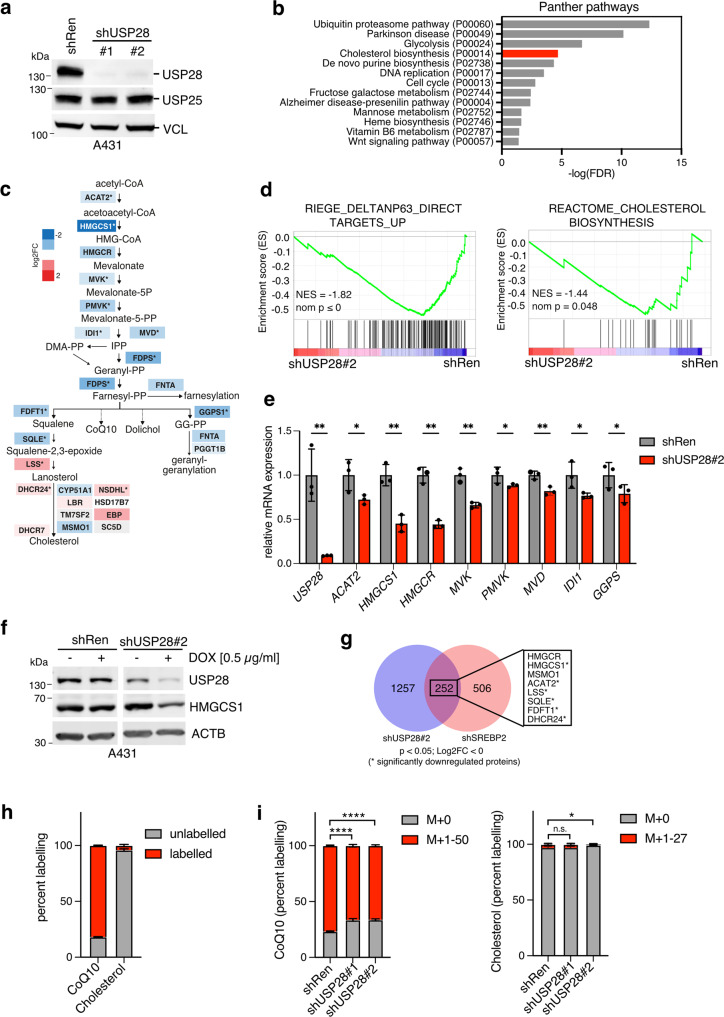
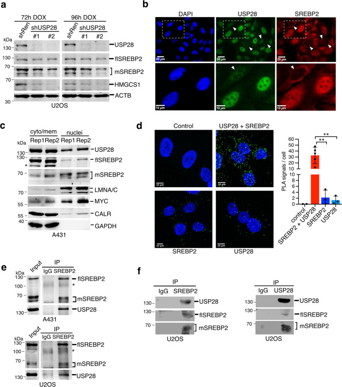
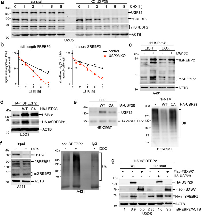
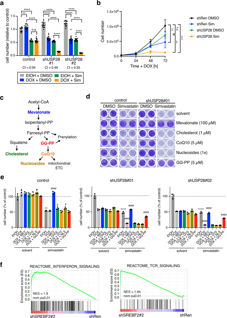
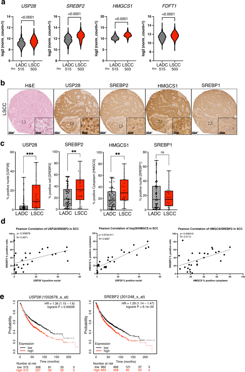
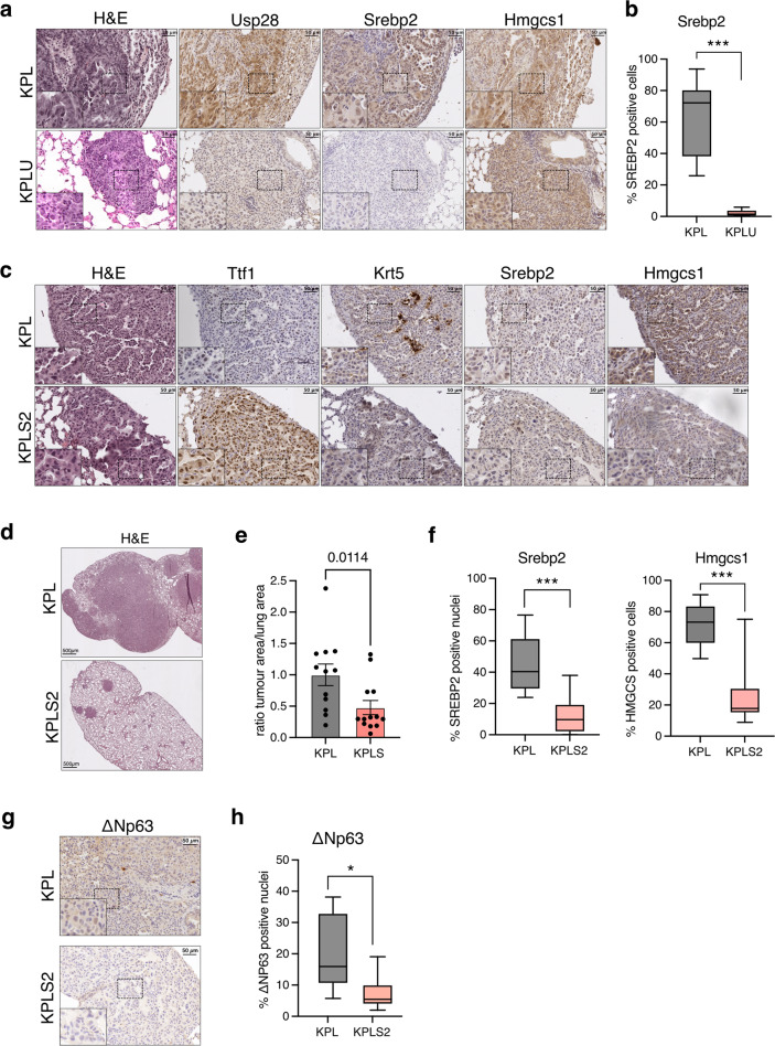
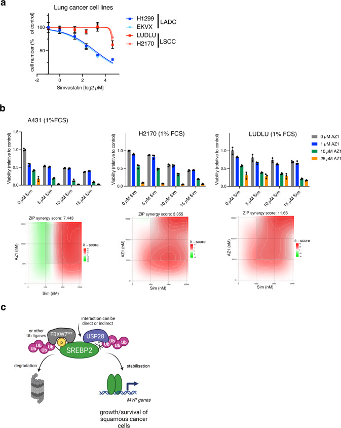
SREBP2 is a master regulator of the mevalonate pathway (MVP), a biosynthetic process that drives the synthesis of dolichol, heme A, ubiquinone and cholesterol and also provides substrates for protein prenylation. Here, we identify SREBP2 as a novel substrate for USP28, a deubiquitinating enzyme that is frequently upregulated in squamous cancers. Our results show that silencing of USP28 reduces expression of MVP enzymes and lowers metabolic flux into this pathway. We also show that USP28 binds to mature SREBP2, leading to its deubiquitination and stabilisation. USP28 depletion rendered cancer cells highly sensitive to MVP inhibition by statins, which was rescued by the addition of geranyl-geranyl pyrophosphate. Analysis of human tissue microarrays revealed elevated expression of USP28, SREBP2 and MVP enzymes in lung squamous cell carcinoma (LSCC) compared to lung adenocarcinoma (LADC). Moreover, CRISPR/Cas-mediated deletion of SREBP2 selectively attenuated tumour growth in a KRas/p53/LKB1 mutant mouse model of lung cancer. Finally, we demonstrate that statins synergise with a dual USP28/25 inhibitor to reduce viability of SCC cells. Our findings suggest that combinatorial targeting of MVP and USP28 could be a therapeutic strategy for the treatment of squamous cell carcinomas.


Conflict of interest statement

The authors declare no competing interests.

Figures

Fig. 1
Fig. 1. USP28 regulates the mevalonate pathway (MVP).
a A431 cells expressing inducible shRNA sequences targeting USP28 (shUSP28#1 and shUSP28#2) or non-targeting control (shRen) were treated with 1 µg/ml of doxycycline (DOX) for 96 h. Protein lysates were analysed for expression of USP28 and USP25 by immunoblotting. Vinculin is shown as loading control. b Proteins differentially regulated by USP28 (shUSP28#1) silencing for 72 h in A431 cells [10] (FDR ≤ 0.05 compared to shRen, total of 2200 proteins) were subjected to pathway analysis using the PANTHER tool. c Differential expression of MVP proteins as determined by proteomics analysis. Pathway map is coloured by log2FC (*q-value ≤ 0.05, n = 3). d A431 cells expressing an inducible shRNA sequence targeting USP28 (shUSP28#2) or non-targeting control (shRen) were treated with 1 µg/ml of doxycycline (DOX) for 120 h. Changes in gene expression were analysed and enrichment plots for gene sets mapping to direct ΔNP63 targets and cholesterol biosynthesis are shown. e Validation of downregulation of mevalonate pathway genes following USP28 silencing using 1 µg/ml of doxycycline (DOX) for 96 h. Data are presented as mean ± SD of three independent experiments (*p < 0.05, ****p < 0.0001, unpaired two-tailed t-test with FDR). f A431 cells expressing an inducible shRNA sequence targeting USP28 (shUSP28#2) or non-targeting control (shRen) were treated with 0.5 µg/ml of doxycycline (DOX) for 96 h. Expression of USP28 and HMGCS1 were analysed by immunoblotting. Actin is shown as loading control. g Overlap between genes downregulated by silencing of USP28 and SREBP2 in A431 cells. Among the 252 overlapping genes are 8 mevalonate pathway enzymes (boxed) that also showed downregulation in the proteomics dataset. h A431 cells were incubated with medium containing 25 mM [U13C]-glucose for 48 h. Metabolites were extracted and analysed by LC-MS. Fractions of labelled and unlabelled ubiquinone (CoQ10) and cholesterol are shown. Data are presented as mean ± SD of three independent experiments. i A431 cells expressing inducible shRNA sequences targeting USP28 (shUSP28#1 and shUSP28#2) or non-targeting control (shRen) were treated with 1 µg/ml of doxycycline (DOX) for 120 h. During the last 48 h, cells were incubated with medium containing 25 mM [U13C]-glucose. Metabolites were extracted and analysed by LC-MS. Fractions of labelled and unlabelled ubiquinone (CoQ10) and cholesterol are shown. Data are presented as mean ± SD of three independent experiments (n.s. non-significant, *p < 0.05, ****p < 0.0001, one-way ANOVA with post-hoc Dunnett’s test).
Fig. 2
Fig. 2. USP28 regulates SREBP2 via close interaction.
a U2OS cells expressing inducible shRNA sequences targeting USP28 (shUSP28#1 and shUSP28#2) or non-targeting control (shRen) were treated with 1 µg/ml of doxycycline (DOX) for 72 or 96 h. Protein lysates were analysed for expression of USP28, full length (flSREBP2) and mature SREBP2 (mSREBP2) and HMGCS1 by immunoblotting. Actin is shown as loading control. b U2OS cells were stained for USP28 and SREBP2 by immunofluorescence. DAPI was used to mark nuclei. Cells with low nuclear expression of USP28 and SREBP2 are marked with arrowheads. c A431 cells were subjected to cell fractionation into cytoplasm and nuclei. Fractions were analysed for expression of USP28 as well as full length (flSREBP2) and mature SREBP2 (mSREBP2). Calreticulin served as marker for ER-membranes, c-Myc and Lamin A/C as markers for nuclei and GAPDH as marker for cytoplasm. Unspecific bands for flSREBP2 are marked by asterisks. Two independent replicate fractionations are shown. d U2OS cells were analysed by proximity ligation assay using antibodies specific for USP28 and SREBP2 either alone or in combination. Number of PLA signals per cell were analysed in >42 cells from at least three biologically independent samples. Data are presented as mean ± SD (*p < 0.05, **p < 0.01, one-way ANOVA with post-hoc Dunnett’s test). e A431 and U2OS cells were subjected to cross-linking with DSP. Cells were fractionated and lysates were subjected to immunoprecipitation using antibodies detecting SREBP2 or matched immunoglobulin controls (IgG). Input and precipitates were analysed for presence of full length and mature SREBP2 as well as USP28. IgG bands are marked by asterisks. f Lysates of U2OS cells were subjected to immunoprecipitation using antibodies detecting SREBP2 (left) or USP28 (right). Precipitates were analysed for presence of full-length and mature SREBP2 as well as USP28.
Fig. 3
Fig. 3. USP28 controls the stability of mature SREBP2.
a U2OS cells were transfected with gRNAs targeting USP28 (KO USP28) and parental controls were treated with 50 µg/ml cycloheximide for the indicated times. Levels of USP28 as well as full length (flSREBP2) and mature SREBP2 (mSREBP2) were determined by immunoblotting. Actin is shown as loading control. b Quantification of full-length and mature SREBP2 signals relative to actin from (a). c A431 cells expressing inducible shRNA sequences targeting USP28 (shUSP28#2) were treated with 0.5 µg/ml of doxycycline (DOX) or solvent (ethanol, EtOH) for 72 h. During the last 6 h, 20 µM MG-132 was added. Cells were lysed and analysed for expression of full-length (flSREBP2) and mature SREBP2 (mSREBP2). Actin is shown as loading control. d U2OS cells were transfected with expression vectors coding for HA-tagged mature SREBP2 (aa 1-484, HA-mSREBP2) together with HA-tagged wild type (WT) or catalytically inactive (CA) USP28. Levels of USP28 and mature SREBP2 were determined by immunoblotting. Actin is shown as loading control. e HEK293 cells were transfected with expression vectors coding for HA-tagged USP28 or HA-tagged mSREBP2 together with His6-Ubiquitin (K48-only). Ubiquitinated proteins were purified using Ni-NTA and analysed by immunoblotting (Ub). Presence of USP28 and SREBP2 was confirmed in the input. f A431 cells expressing inducible shRNA sequences targeting USP28 (shUSP28#2) were treated with 1 µg/mL doxycycline (DOX) or solvent (ethanol) for 96 h with 20 µM MG132 being added during the last 6 h. Cells were lysed and subjected to immunoprecipitation using antibodies detecting SREBP2. Precipitates were analysed for the presence of ubiquitinated proteins (Ub). Efficient silencing of USP28 and presence of full length and mature SREBP2 was confirmed in the input. Actin is shown as a loading control. IgG bands are marked by asterisks. g U2OS cells were transfected with either wild type or CPD mutant (Ser 432 and 436 to Ala) HA-tagged mature SREBP2 together with HA-tagged USP28 or Flag-FBXW7. Levels of USP28, FBXW7 and mature SREBP2 were determined by immunoblotting. Actin is shown as loading control.
Fig. 4
Fig. 4. Inhibition of USP28 and MVP synergise in reducing cell viability.
a A431 cells expressing inducible shRNAs targeting USP28 (shUSP28#1 or shUSP28#2) or control cells were treated with 1 µg/ml doxycycline (Dox) or solvent (ethanol, EtOH) for 72 h together with 10 µM simvastatin (Sim) or solvent (DMSO). Cell viability was determined by crystal violet staining. Data are presented as mean± SD of three independent experiments (n.s. non-significant, ****p < 0.00001, one-way ANOVA with post-hoc Tukey test). b Growth curves of shUSP28#1 or control (shRen) treated with 1 µg/ml doxycycline (Dox) or solvent (ethanol, EtOH) together with 10 µM simvastatin (Sim) or solvent (DMSO). Data are presented as mean± SD of three independent experiments (n.s. non-significant, *p < 0.05, ****p < 0.00001, one-way ANOVA with post-hoc Tukey test). c Schematic of the different metabolic outputs of the MVP. GG-PP = geranyl-geranyl-pyrophosphate, CoQ10 = ubiquinone. d Cells as in (a) were treated with 1 µg/ml doxycycline or solvent (ethanol, EtOH) for 72 h together with 10 µM simvastatin (Sim) or solvent (DMSO) plus the indicated compounds: 100 µM mevalonate, 1 µM cell-permeable cholesterol, 5 µM ubiquinone (CoQ10), nucleosides (30 µM C, G, A, U each; 10 µM T) or 5 µM GG-PP. Cell viability was determined by crystal violet staining. Representative images of three independent replicates are shown. e Quantitation of data shown in (d). Data are presented as mean± SD of three independent replicates (n.s. non-significant, **p < 0.01, ****p < 0.0001, unpaired two-tailed Student’s t-test between EtOH vs EtOH plus simvastatin; #p < 0.05, ####p < 0.0001, one-way ANOVA with post-hoc Dunnett’s test compared to DOX plus simvastatin). f GSEA enrichment plots for gene sets mapping to interferon and TCR signalling comparing cells depleted for SREBF2 (SREBF2#2) and control (shRen).
Fig. 5
Fig. 5. SREBP2 is upregulated in squamous cell carcinoma.
a Violin plots showing expression of USP28, SREBF2, HMGCS1 and FDFT1 in human Lung adenocarcinoma (ADC) and lung squamous carcinoma (SCC). Data are from TCGA (Firehose legacy). Significance was calculated using Mann–Whitney test. ****p < 0.0001. b Tissue sections from a human NSCLC TMA were stained for USP28, SREBP2 HMGCS1 and SREBP1 by immunohistochemistry. Haematoxilin and eosin (H&E) staining is also shown. Representative images for SCC are shown. c Boxplots showing quantification of USP28, SREBP2, HMGCS and SREBP1 staining in SCC and ADC tumours. Percent positive cells are shown. (SCC: n = 33; ADC: n = 75; ***p < 0.001, Mann-Whitney test). d Pearson correlation between expression of USP28, SREBP2 and HMGCS in SCC tumours (R = Pearson correlation coefficient, p = two tailed t-test). e Kaplan–Meier plots of overall survival of lung cancer patients (N = 1925) dichotomised into ‘high’ and ‘low’ based on median USP28 or SREBF2 expression score. Survival differences were calculated with the log-rank test.
Fig. 6
Fig. 6. Deletion of Srebf2 attenuates tumour formation in a mouse model of lung squamous cell carcinoma.
a Tissue sections from KPL and KPLU lung tumours were stained for Usp28, Srebp2 and Hmgcs1 by immunohistochemistry. Haematoxylin and eosin (H&E) staining is also shown. b Boxplot showing quantification of Srebp2 staining in KPL and KPLU tumours. Percent positive cells are shown. (KPL: n = 9; KPLU: n = 7; ***p < 0.001, Mann–Whitney test). c Tissue sections from KPL and KPLS2 tumours were stained for the adenocarcinoma marker thyroid transcription factor 1/NK2-homeobox 1 (Ttf-1/Nkx2-1), the squamous marker keratin 5 (Krt5), Srebp2 and Hmgcs1 by immunohistochemistry. H&E staining is also shown. d H&E staining of representative lung tissue sections from KPL and KPLS2 mice. e Ratio of tumour area relative to total lung area in KPL and KPLS2 mice. Data are displayed as mean ± SD (KPL: n = 11; KPLS2: n = 13; Mann-Whitney test). f Boxplot showing quantification of Srebp2 and Hmgcs1 staining in KPL and KPLS2 tumours. Percent positive nuclei or positive cells are shown. (KPL: n = 7; KPLS2: n = 9; ***p < 0.001, Mann-Whitney test). g Tissue sections from KPL and KPLS2 tumours were stained for ΔNp63. h Boxplot showing quantification of ΔNp63 staining in KPL and KPLS2 tumours. Percent positive nuclei are shown. (KPL: n = 6; KPLS2: n = 6; ***p < 0.05, Mann-Whitney test).
Fig. 7
Fig. 7. Simvastatin synergises with a dual USP28/25 inhibitor to reduce viability of SCC cells.
a Human ADC (H1299 and EKXV) and SCC (LUDLU and H2170) cells were treated with different concentrations of simvastatin for 72 h. Cell viability was determined using crystal violet staining. b A431, H2170 and LUDLU cells were treated with indicated concentrations of simvastatin (Sim) or AZ1 in medium containing 1% FCS for 72 h. Drug synergy was calculated using ZIP model. c Working model for the regulation of SREBP2 by USP28. Created with BioRender.com.

References

    1. Oh E, Akopian D, Rape M. Principles of Ubiquitin-Dependent Signaling. Annu Rev Cell Dev Biol. 2018;34:137–62. doi: 10.1146/annurev-cellbio-100617-062802. - DOI - PubMed
    1. Komander D, Rape M. The ubiquitin code. Annu Rev Biochem. 2012;81:203–29. doi: 10.1146/annurev-biochem-060310-170328. - DOI - PubMed
    1. Mevissen TET, Komander D. Mechanisms of Deubiquitinase Specificity and Regulation. Annu Rev Biochem. 2017;86:159–92. doi: 10.1146/annurev-biochem-061516-044916. - DOI - PubMed
    1. Sacco JJ, Coulson JM, Clague MJ, Urbe S. Emerging roles of deubiquitinases in cancer-associated pathways. IUBMB Life. 2010;62:140–57. - PMC - PubMed
    1. Muller I, Strozyk E, Schindler S, Beissert S, Oo HZ, Sauter T, et al. Cancer Cells Employ Nuclear Caspase-8 to Overcome the p53-Dependent G2/M Checkpoint through Cleavage of USP28. Mol Cell. 2020;77:970–84.e7. doi: 10.1016/j.molcel.2019.12.023. - DOI - PMC - PubMed

Publication types

Substances