Choroidal lesions in varicella zoster virus uveitis
- PMID: 37203072
- PMCID: PMC10391431
- DOI: 10.4103/ijo.IJO_2099_22
Choroidal lesions in varicella zoster virus uveitis
Abstract
Purpose: To evaluate choroidal lesions with spectral domain optical coherence tomography (SD-OCT) scan in varicella zoster virus (VZV) uveitis.
Methods: VZV-uveitis cases which underwent OCT scan for choroidal lesions were studied. SD-OCT scan passing through these lesions was studied in detail. Subfoveal choroidal thickness (SFCT) during active and resolved stages was studied. Angiogaphic features were studied where available.
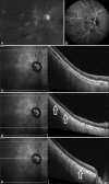
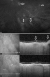
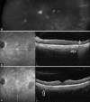
Results: Thirteen out of 15 cases had same-sided herpes zoster ophthalmicus skin rashes. All except three patients had old or active kerato-uveitis. All eyes demonstrated clear vitreous and a single or multiple hypopigmented orangish-yellow choroidal lesions. The number of lesions remained unchanged during the follow-up on clinical examination. SD-OCT over lesions (n = 11) showed choroidal thinning (n = 5), hyporeflective choroidal elevation during active inflammation (n = 3), transmission effects (n = 4), and ellipsoid zone disruption (n = 7). The mean change in SFCT (n = 9) after resolution of the inflammation was 26.3 μm (range: 3-90 μm). Fundus fluorescein angiography showed iso-fluorescence over lesions in all (n = 5), but indocyanine green angiography (n = 3) showed hypofluorescence at lesions. Mean follow-up was 1.38 years (range: 3 months-7 years). De-novo appearance of choroidal lesion during the first relapse of VZV-uveitis was captured in one case.
Conclusion: VZV-uveitis can cause focal or multifocal hypopigmented choroidal lesions with thickening or scarring of choroidal tissue, depending on the disease activity.
Keywords: Choroidal granuloma; FFA; ICG; OCT; OCTA; VZV; choroidal vitiligo; choroiditis; herpes; hypopigmented choroidal lesion; varicella zoster.
Conflict of interest statement
None
Figures


References
-
- Babu K, Mahendradas P, Sudheer B, Kawali A, Parameswarappa DC, Pal V, et al. Clinical profile of herpes zoster ophthalmicus in a South Indian patient population. Ocul Immunol Inflamm. 2018;26:178–83. - PubMed
-
- Puri LR, Shrestha GB, Shah DN, Chaudhary M, Thakur A. Ocular manifestations in herpes zoster ophthalmicus. Nepal J Ophthalmol. 2011;3:165–71. - PubMed
-
- Singh J, Bhatia P, Sen A, More A. Choroidal involvement in a case of acute retinal necrosis. Ocul Immunol Inflamm. 2022;16:1–5. - PubMed
-
- Meenken C, Rothova A. Varicella-Zoster virus–associated multifocal chorioretinitis in 2 boys. JAMA Ophthalmol. 2013;131:969–70. - PubMed
MeSH terms
LinkOut - more resources
Full Text Sources

