. 2023 Dec;41(12):2599-2609.
doi: 10.1002/jor.25627. Epub 2023 May 25.

Development and expansion of intramuscular adipose tissue is not dependent on UCP-1-lineage cells in mice

Affiliations

Development and expansion of intramuscular adipose tissue is not dependent on UCP-1-lineage cells in mice

Jacob C Parson et al. J Orthop Res. 2023 Dec.

Abstract

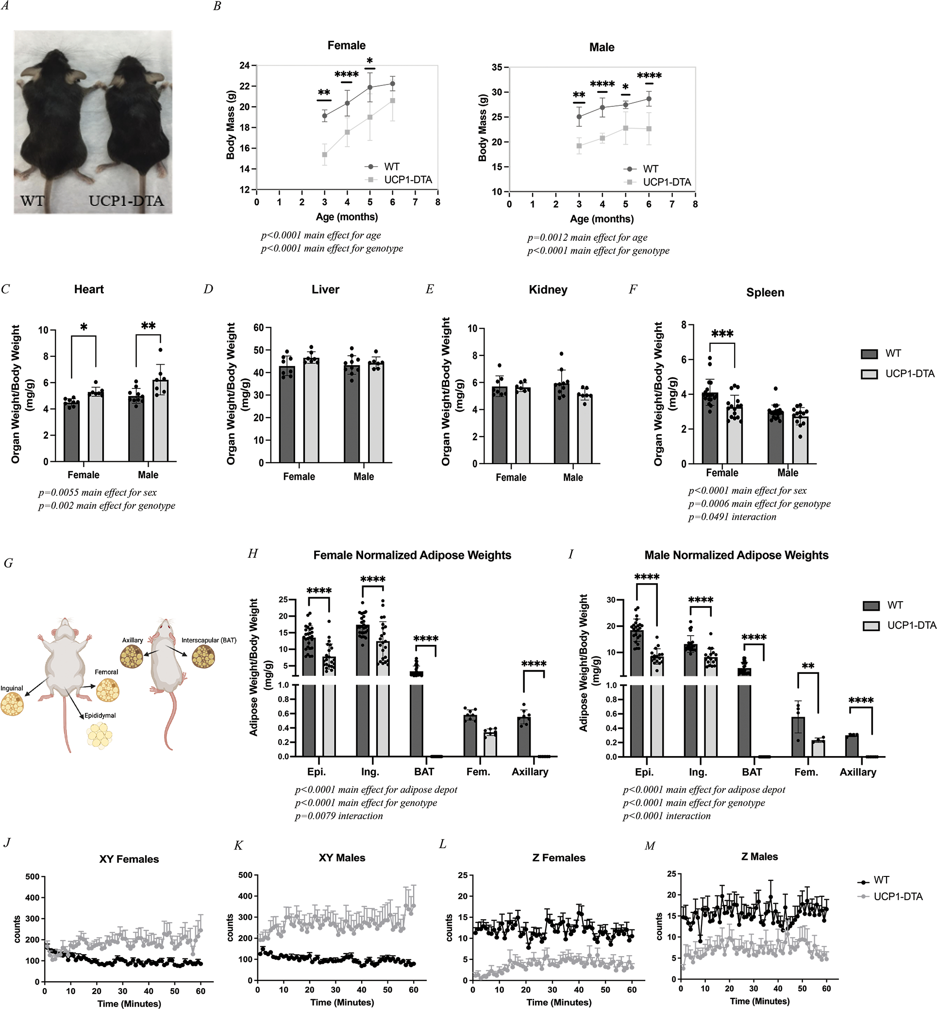
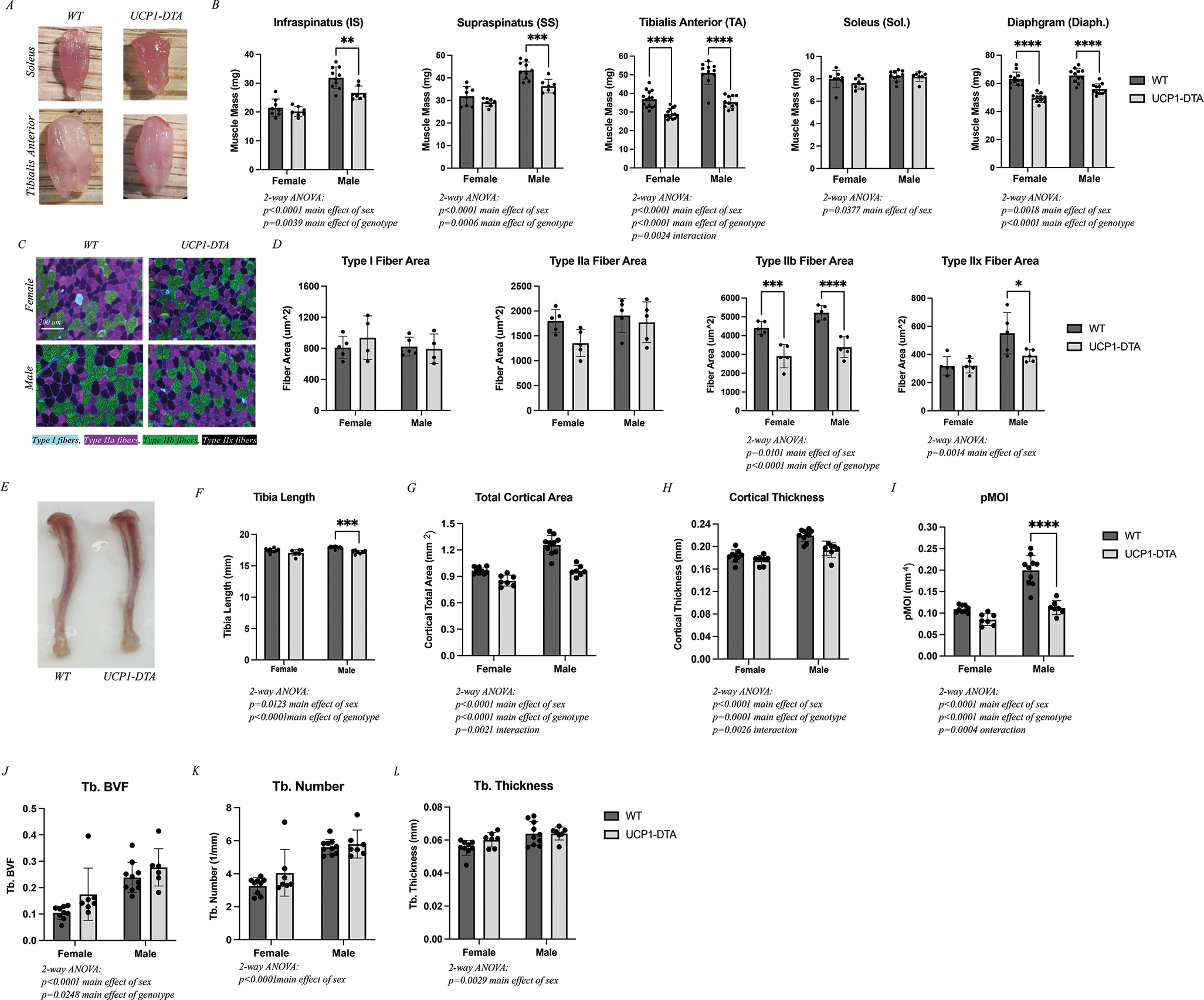
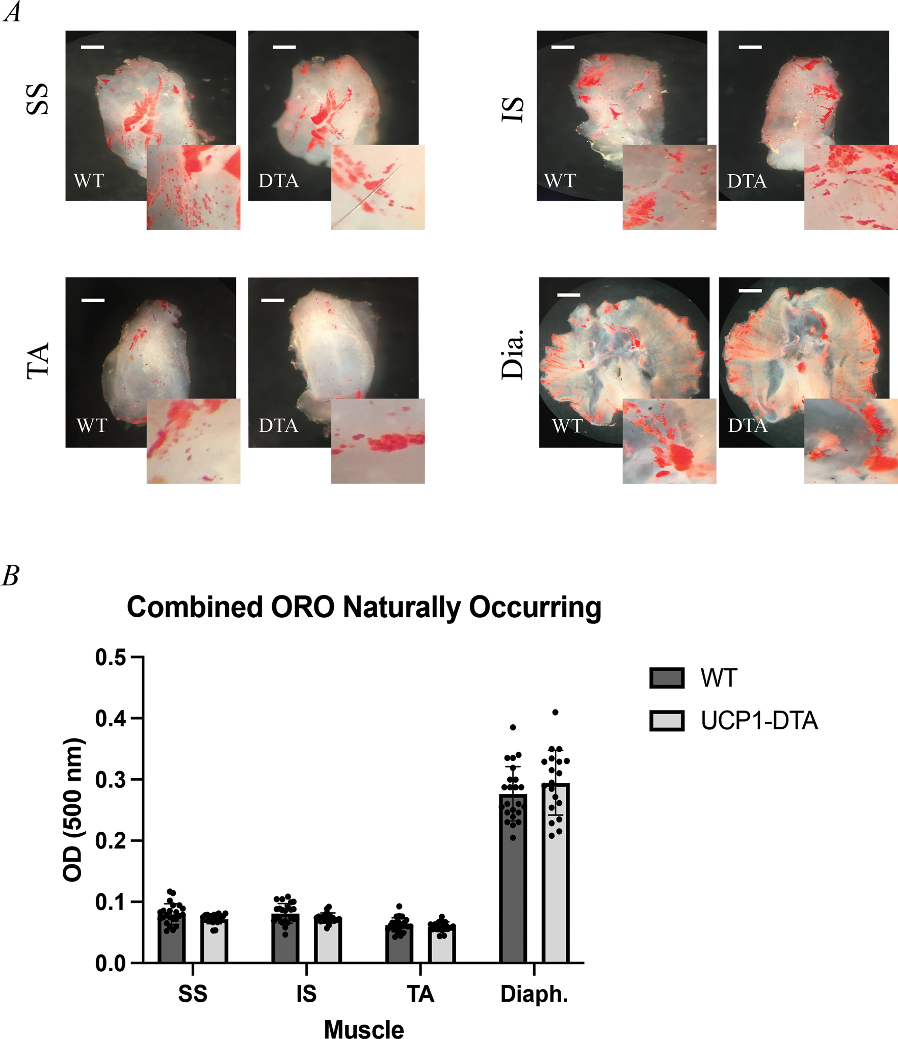
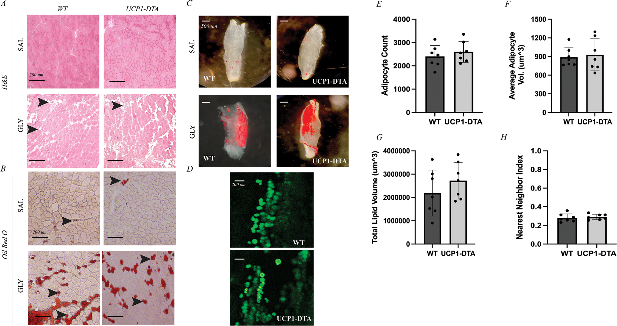
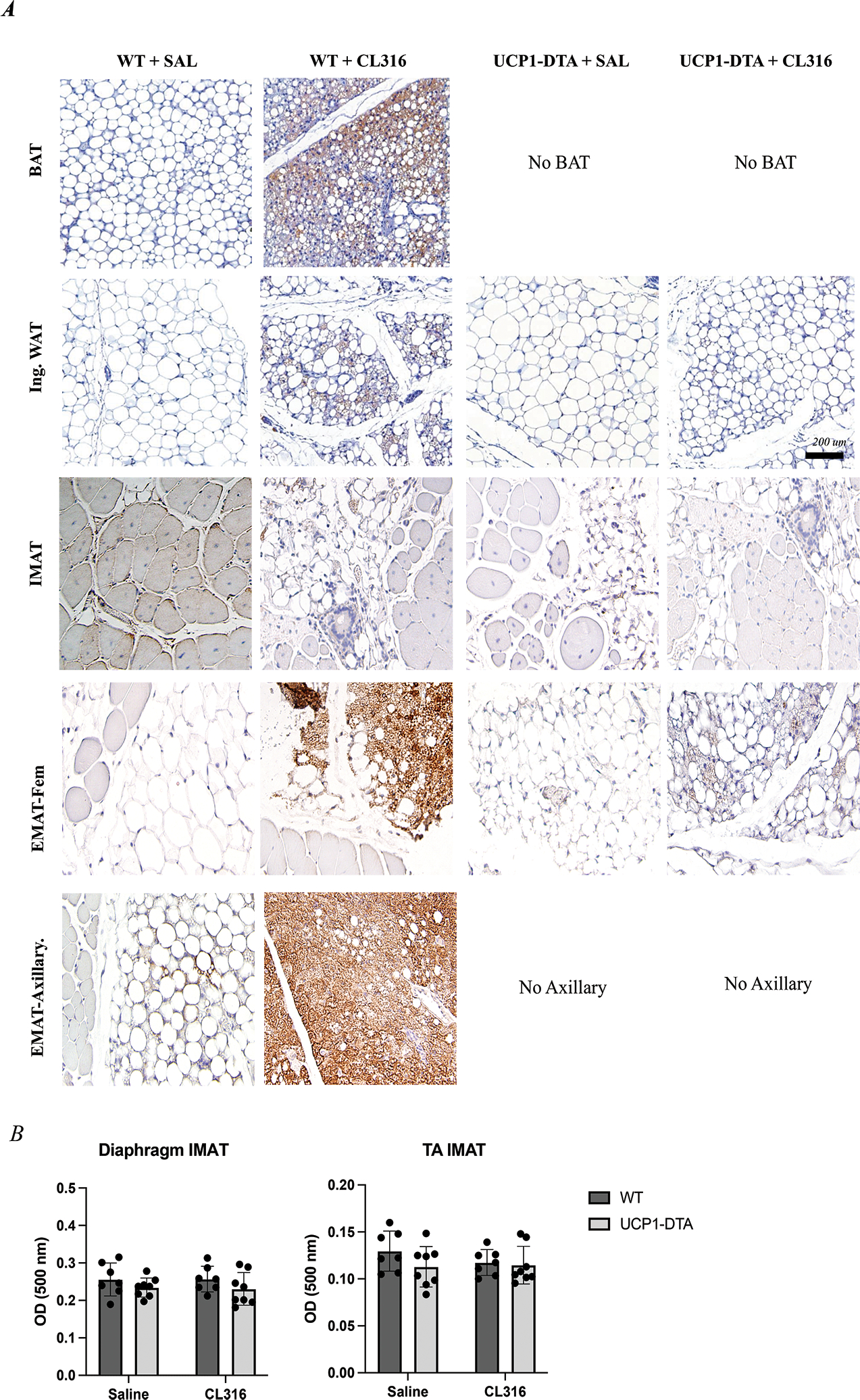
Accumulation of adipose tissue within and outside of skeletal muscle is associated with orthopedic injury and metabolic disease, where it is thought to impede muscle function. The close juxtaposition between this adipose and myofibers has led to hypotheses that paracrine interactions between the two regulate local physiology. Recent work suggests that intramuscular adipose tissue (IMAT) may have features of beige or brown fat, indicated by the expression of uncoupling protein-1 (UCP-1). However, this is contested by other studies. Clarification of this point is needed to inform our understanding of the relationship between IMAT and muscle health. To achieve this, we examined the effects of constitutive UCP-1+ cell ablation (UCP1-DTA) on IMAT development and homeostasis. IMAT developed normally in UCP1-DTA mice, with no significant differences in quantity compared with wild-type littermates. Likewise, IMAT accumulation in response to glycerol-induced injury was similar between genotypes, with no significant differences in adipocyte size, quantity, or dispersion. This suggests that neither physiological nor pathological IMAT express UCP-1 and that the development of IMAT does not depend on UCP-1 lineage cells. In response to β3-adrenergic stimulation, we find minor, localized UCP-1 positivity in wildtype IMAT, but the bulk of the adipocytes are unresponsive. In contrast, two depots of muscle-adjacent (epi-muscular) adipose tissue have reduced mass in UCP1-DTA mice and UCP-1 positivity in wildtype littermates, comparable to traditional beige and brown adipose depots. Taken together this evidence strongly supports a white adipose phenotype for mouse IMAT and a brown/beige phenotype for some adipose outside the muscle boundary.

Keywords: muscle; pathophysiology; progenitors and stem cells.


Figures

Figure 1.
Figure 1.. UCP1-DTA Mice Have Reduced Body and Adipose Masses
A Representative image illustrating body size differences between WT mouse (left) and UCP1-DTA mouse (right). B Body mass was significantly lower in UCP1-DTA mice across cohort ages. C Normalized heart mass is significantly higher in UCP1-DTA mice. D-E Liver and kidney analyses show no differences between sex or genotype. F Spleen mass was significantly reduced in female mice only. G Graphical anatomical depiction of six adipose depots including three classical fat depots: Epididymal (Epi.), Inguinal (Ing.), and interscapular (BAT) and two previously identified epi-muscular fat depots including Femoral (Fem.) and Axillary adipose tissue. H Normalized adipose masses in female mice show main effects for adipose depot and genotype with specific differences noted between Epi, Ing., BAT, and Axillary depots. I Normalized adipose masses in male mice demonstrate main effects for adipose depot and genotype with specific differences noted across all five fat depots between genotypes. J-M Activity data in WT and UCP1-DTA female and male mice shows that in XY directions, UCP1-DTA mice are significantly more active than WT mice, but significantly less active in the Z direction. Data are presented as mean ± SD. *P<0.05, **P<0.01, ***P<0.001, ****P<0.0001. Statistical significance set at p<0.05. Analyses were in Prism using two-way ANOVA or multiple, independent t-tests, or linear regression.
Figure 2.
Figure 2.. UCP1-DTA Mice Show Altered Skeletal Muscle and Bone Growth
A Images of WT and UCP1-DTA soleus and tibialis anterior (TA) muscles illustrate gross size differences in the TA, but not the soleus. B Raw muscle masses harvested from female and male WT and UCP1-DTA mice including infraspinatus (IS), supraspinatus (SS), tibialis anterior (TA), soleus (Sol.), and diaphragm (Diaph.) demonstrating reduced mass in all but soleus muscles. C Representative 20x images of TA muscles from both female and male WT and UCP1-DTA immunostained against myosin heavy chain isoforms to detect specific fiber types. Type I fibers: Cyan, Type IIa: Magenta, Type IIb: Green, and Type IIx: Black/No channel. Scale bar represents 200 μm. D Analysis of fiber area by fiber type found a significant reduction in type IIb fiber area in UCP1-DTA males and females and type IIx fibers in males only. E Representative tibia bone images illustrate a small decrease in length in UCP1-DTA mice. F Analysis of tibia length with male WT mice have significantly longer tibias than UCP1-DTA males. G-I Total cortical area, cortical thickness and pMOI are modestly reduced in UCP1-DTA mice compared with WT. J-L Ttrabecular (Tb.) bone volume fraction (BVF), Tb. Number and Tb. Thickness are similar between UCP1-DTA and WT male and female mice. N=8–12. Data are presented as mean ± SD. *P<0.05, **P<0.01, ***P<0.001, ****P<0.0001. Statistical significance set at p<0.05. Analyses were in Prism using two-way ANOVA or multiple, independent t-tests.
Figure 3.
Figure 3.. Intramuscular Adipose Tissue (IMAT) Develops Without UCP-1-Expressing Cells
Oil Red O staining of decellularized A Supraspinatus (SS), Infraspinatus (IS), Tibialis Anterior (TA), and Diaphragm (Dia.) muscles depicting visually similar amounts of naturally occurring IMAT (red) in both wild type (WT) and UCP1-DTA genotypes. B Optical density (OD) measurement confirms similar amount of lipid content across the four muscles in both female (n=14) and male (n=13) mice. Scale bar 1mm. Analysis performed in Prism using multiple, independent t-tests. Statistical significance set at p<0.05.
Figure 4.
Figure 4.. Glycerol-Induced IMAT Develops Without UCP-1-Expressing Cells
A Hematoxylin and Eosin (H&E) staining of tibialis anterior (TA) muscle after 14 days of either saline (SAL) or glycerol (GLY) treatment shows extensive appearance of intramuscular adipocytes (IMAT) in GLY groups represented by white, globular structures (black arrows). B Oil Red O (ORO) staining of TA muscles identifies these intramuscular adipocytes as lipid filled (black arrows). C Oil Red O staining of decellularized EDL muscles of both wild type (WT) and UCP1-DTA genotypes injected with saline showing minimal IMAT (red) or injected with glycerol causing IMAT accumulation in similar amount between genotypes (red). Scale bars for A and B 1 mm. D Sample slices of EDLs treated with glycerol and stained with BODIPY and visualized using confocal microscopy in WT and UCP1-DTA mice. Individual adipocytes are identified as individual green circular regions of interest. E Quantification of adipocyte metrics via confocal microscopy in male WT and UCP1-DTA mice (n=21) showing similar average adipocyte count between genotypes in response to glycerol injection. F Average adipocyte volume similar in WT and UCP1-DTA mice. G Total lipid volume of WT and UCP1-DTA mice are similar with glycerol injection. H Nearest neighbor index as a measure of spatial clustering of intramuscular adipocytes show similar clustering between WT and UCP1-DTA mice. Data are presented as mean ± SD. Analysis performed in Prism using independent t-tests. Statistical significance set at p<0.05.
Figure 5.
Figure 5.. Effects of Beiging on Adipose Depots
A) Representative images from UCP-1-immunostained adipose depots Row 1 In WT micce, brown adipose (BAT) adipocytes have multilocular morphology, and increased UCP-1 immunostaining intensity with CL316,243 (β3 adrenergic agonist) treatment (brown). UCP1-DTA mice completely lack the classical interscapular, BAT depot. Row 2 Inguinal white adipose (Ing. WAT) has interspersed unilocular and multilocular adipocytes in saline (SAL) treated mice with increased UCP-1+ multilocular cells following CL316 treatment in WT, but less so in UCP1-DTA. Row 3 Tibialis anterior (TA) muscles of both WT and UCP1-DTA genotypes injected with glycerol show deposition of IMAT across all genotypes and conditions noted by unilocular adipocytes. Both WT and UCP1-DTA mice treated with CL316 show small localized UCP-1+ cells, however, to a much lesser extent than Ing. WAT. Row 4 Femoral Epi-muscular adipose depots (Fem. EMAT) from WT and UCP1-DTA mice show predominantly large, unilocular lipid droplets in saline-treated conditions. WT and UCP1-DTA Fem. EMAT treated with CL316 show a dramatic increase in the intensity of UCP-1 immunostaining. Row 5 Similar to Ing. WAT, Axillary EMAT have interspersed unilocular and multilocular adipocytes in saline-treated condition, and when treated with CL316 have dramatically higher UCP-1 staining intensity. B There are no changes in total IMAT quantity in response to CL316 treatment in either diaphragm (Diaph.) or glycerol-treated TA in response to CL316. N= 8–19. Data are presented as mean ± SD. *P<0.05, **P<0.01, ***P<0.001, ****P<0.0001. Statistical significance set at p<0.05. Analyses were in Prism using two-way ANOVA. Scale bar is 200 μm.

References

    1. Giralt M, Villarroya F. 2013. White, brown, beige/brite: different adipose cells for different functions? Endocrinology 154:2992–3000. - PubMed
    1. Villarroya F, Cereijo R, Villarroya J, et al. 2017. Brown adipose tissue as a secretory organ. Nat Rev Endocrinol 13:26–35. - PubMed
    1. Kajimura S, Spiegelman BM, Seale P. 2015. Brown and Beige Fat: Physiological Roles beyond Heat Generation. Cell Metab 22:546–559. - PMC - PubMed
    1. Addison O, Marcus RL, Lastayo PC, et al. 2014. Intermuscular fat: a review of the consequences and causes. Int J Endocrinol 2014:309570. - PMC - PubMed
    1. Wang H, Willershäuser M, Karlas A, et al. 2019. A dual Ucp1 reporter mouse model for imaging and quantitation of brown and brite fat recruitment. Mol Metab 20:14–27. - PMC - PubMed

Publication types

Substances