

Linocin M18 protein from the insect pathogenic bacterium Brevibacillus laterosporus isolates
- PMID: 37204448
- PMCID: PMC10313851
- DOI: 10.1007/s00253-023-12563-8
Linocin M18 protein from the insect pathogenic bacterium Brevibacillus laterosporus isolates
Abstract
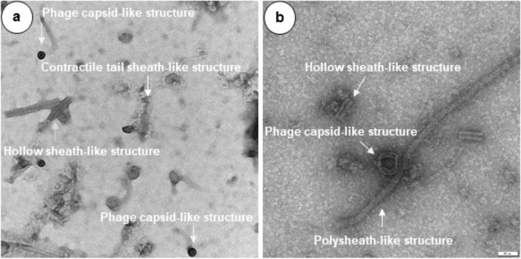
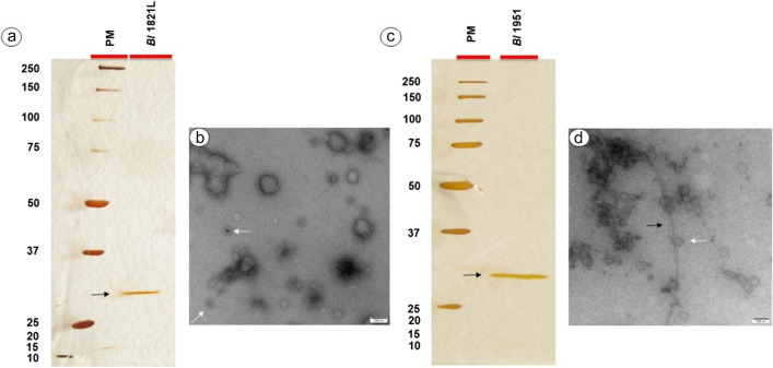
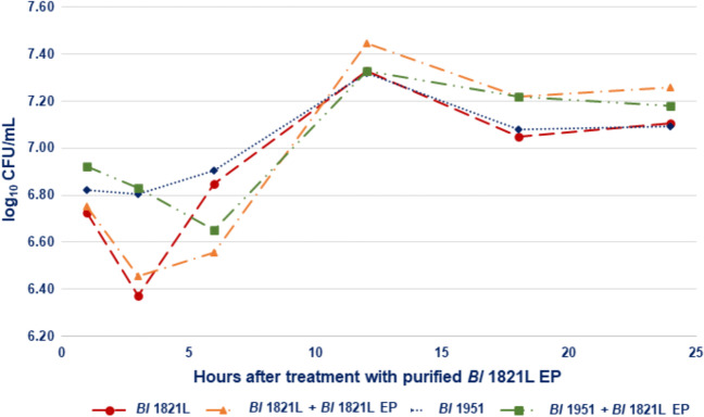
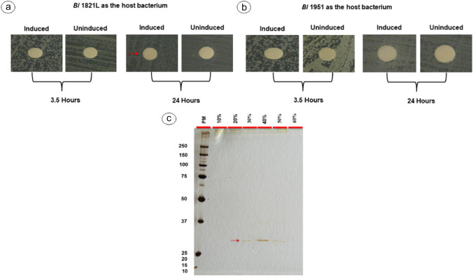
Brevibacillus laterosporus (Bl) is a Gram-positive and spore-forming bacterium. Insect pathogenic strains have been characterised in New Zealand, and two isolates, Bl 1821L and Bl 1951, are under development for use in biopesticides. However, growth in culture is sometimes disrupted, affecting mass production. Based on previous work, it was hypothesised that Tectiviridae phages might be implicated. While investigating the cause of the disrupted growth, electron micrographs of crude lysates showed structural components of putative phages including capsid and tail-like structures. Sucrose density gradient purification yielded a putative self-killing protein of ~30 kDa. N-terminal sequencing of the ~30 kDa protein identified matches to a predicted 25 kDa hypothetical and a 31.4 kDa putative encapsulating protein homologs, with the genes encoding each protein adjacent in the genomes. BLASTp analysis of the homologs of 31.4 kDa amino acid sequences shared 98.6% amino acid identity to the Linocin M18 bacteriocin family protein of Brevibacterium sp. JNUCC-42. Bioinformatic tools including AMPA and CellPPD defined that the bactericidal potential originated from a putative encapsulating protein. Antagonistic activity of the ~30 kDa encapsulating protein of Bl 1821L and Bl 1951during growth in broth exhibited bacterial autolytic activity. LIVE/DEAD staining of Bl 1821L cells after treatment with the ~30 kDa encapsulating protein of Bl 1821L substantiated the findings by showing 58.8% cells with the compromised cell membranes as compared to 37.5% cells in the control. Furthermore, antibacterial activity of the identified proteins of Bl 1821L was validated through gene expression in a Gram-positive bacterium Bacillus subtilis WB800N. KEY POINTS: • Gene encoding the 31.4 kDa antibacterial Linocin M18 protein was identified • It defined the autocidal activity of Linocin M18 (encapsulating) protein • Identified the possible killing mechanism of the encapsulins.
Keywords: Antibacterial protein; Brevibacillus laterosporus; Encapsulating protein; Heterologous expression; Insect pathogenic bacterium; Linocin M18 protein.
© 2023. The Author(s).
Conflict of interest statement
The authors declare no competing interests.
Figures


References
-
- Åberg A, Nordlund P, Eklund H. Unusual clustering of carboxyl side chains in the core of iron-free ribonucleotide reductase. Nature. 1993;361(6409):276–278. - PubMed
-
- Akita F, Chong KT, Tanaka H, Yamashita E, Miyazaki N, Nakaishi Y, Suzuki M, Namba K, Ono Y, Tsukihara T. The crystal structure of a virus-like particle from the hyperthermophilic archaeon Pyrococcus furiosus provides insight into the evolution of viruses. J Mol Biol. 2007;368(5):1469–1483. - PubMed
-
- Almeida AV, Carvalho AJ, Pereira AS. Encapsulin nanocages: protein encapsulation and iron sequestration. Coord Chem Rev. 2021;448:214188. doi: 10.1016/j.ccr.2021.214188. - DOI
-
- Al-Sammak EG. Identification of bacteriocin Linocin M18 from Brevibacterium and related genera using PCR. J Biotech Res Center. 2014;8(2):28–33.
-
- Anderson I, Sorokin A, Kapatral V, Reznik G, Bhattacharya A, Mikhailova N, Burd H, Joukov V, Kaznadzey D, Walunas T. Comparative genome analysis of Bacillus cereus group genomes with Bacillus subtilis. FEMS Microbiol Lett. 2005;250(2):175–184. - PubMed
MeSH terms
Substances
Supplementary concepts
LinkOut - more resources
Full Text Sources
