Review
. 2023 Aug;48(8):2705-2715.
doi: 10.1007/s00261-023-03939-5. Epub 2023 May 19.

Abdominal visceral tuberculosis: a malignancy mimic

Affiliations
Review

Abdominal visceral tuberculosis: a malignancy mimic

Chandan J Das et al. Abdom Radiol (NY). 2023 Aug.

Abstract

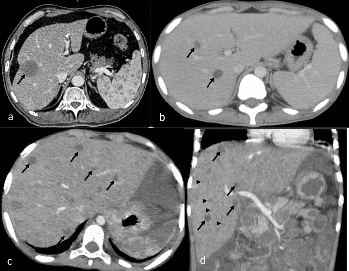
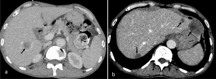
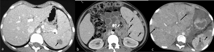
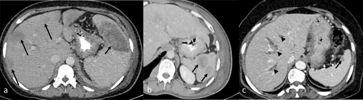
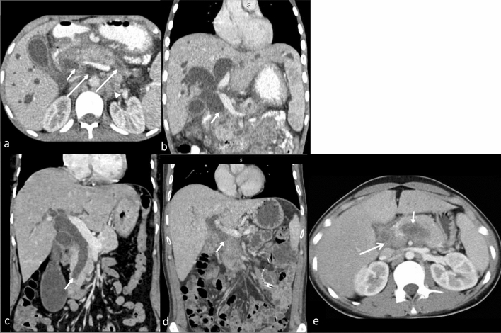
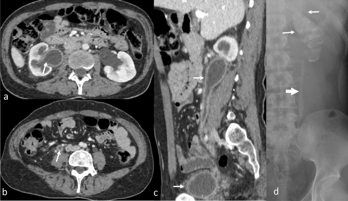
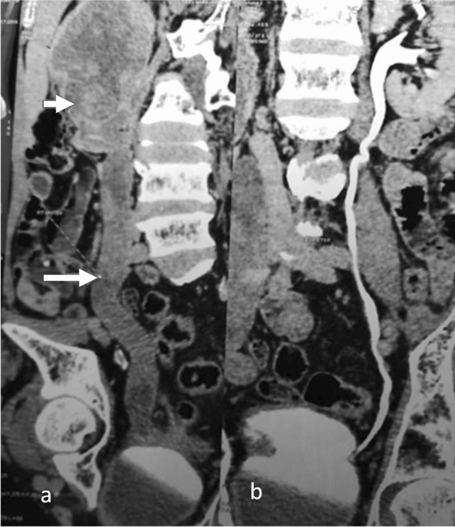
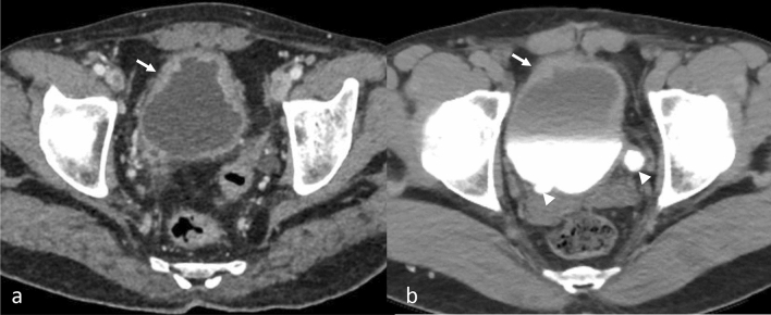
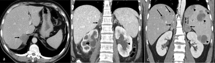
The purpose is to discuss abdominal tuberculosis mimicking malignancy involving the abdominal viscera. TB of the abdominal viscera is common, especially in countries where tuberculosis is endemic and in pockets of non-endemic countries. Diagnosis is challenging as clinical presentations are often non-specific. Tissue sampling may be necessary for definitive diagnosis. Awareness of the early and late disease imaging appearances of abdominal tuberculosis involving the viscera that can mimic malignancy can aid detecting TB, providing a differential diagnosis, assessing extent of spread, guiding biopsy, and evaluating response.

Keywords: Abdominal viscera; CT; Cancer; MRI; Tuberculosis; Ultrasonography.


Conflict of interest statement

The authors have no competing interests to declare that are relevant to the content of this article.

Figures

Fig. 1
Fig. 1
CECT axial images of abdomen show different pattern of liver involvement in various cases of TB (arrow) as (a) large focal low attenuation lesion, (b) few hypodense nodules and, (c, d) hepatomegaly with multiple hypodense nodules, including 0.5–2 mm nodules that can be seen with the micronodular type (arrowhead)
Fig. 2
Fig. 2
CECT abdomen axial image (a) shows a solitary hypoenhancing focal lesion (arrow) in liver with “target” appearance of peripheral enhancement in a known case of rectal cancer suggesting liver metastasis. CECT axial abdomen image (b) shows similar multiple hypoenhancing lesions (arrows) in a patient with colorectal carcinoma, suggesting liver metastases
Fig. 3
Fig. 3
CECT axial images of abdomen show splenic involvement in various cases of TB as (a) large focal lesion with rim enhancement (arrow), splenomegaly with multiple small focal lesions (arrow) (b), and large splenic abscesses (c). Note retroperitoneal adenopathy (thick arrow) in (b) and liver lesions (long arrow) in (c)
Fig. 4
Fig. 4
CECT abdomen images showing various forms of splenic metastases which can mimic TB involvement: (a) large low attenuation splenic metastasis (arrow) and liver metastases (long arrow) from breast cancer, (b) enhancing lesions in the spleen due to non-Hodgkin’s lymphoma, (c) solitary low attenuation splenic metastasis from breast cancer. Intrahepatic ductal dilatation is also seen (arrowheads)
Fig. 5
Fig. 5
CECT abdomen images (a axial, b cor) in a case of TB reveal dilated bilateral intrahepatic biliary radicles and CBD with its abrupt cut off (arrow). Pancreas is bulky with hypodense area in tail and uncinated process (long arrow). Necrotic retroperitoneal nodes (arrowhead) are noted. Similar appearance can be seen with (c, cor) distal cholangiocarcinoma (arrow) and (d, cor) pancreatic adenocarcinoma showing abrupt cut off of the dilated common bile duct. Axial CT (e) image showing a hypodense lesion (short arrow) in pancreatic body with necrotic peri pancreatic node (long arrow) highly suspicious for pancreatic malignancy. On biopsy, it was found to be pancreatic tuberculosis
Fig. 6
Fig. 6
Genitourinary TB (arrow), CECT axial (a, b), and sagittal (c) images depict dilatation, irregular thickening and enhancement of (a) right pelvicalyceal system, (b, c) ureter, and (c) urinary bladder. These findings can be confused with urothelial carcinoma. Plain radiograph (d) shows lobar-pattern of calcification of the entire left kidney signifying putty kidney (white arrows) and calcification of the left upper ureter (block arrow) in an advanced case of TB
Fig. 7
Fig. 7
CECT coronal images of the abdomen in a patient with urothelial carcinoma shows (a) a mass in right pelvis (arrow) and ureter with no contrast excretion (long arrow) whereas (b) normal contrast excretion is seen in the pelvicalyceal system and ureter on the opposite side
Fig. 8
Fig. 8
CECT pelvis axial images (a) show irregular wall thickening of the urinary bladder (arrow) in a patient suspected for urothelial carcinoma who underwent cystoscopy and biopsy. Biopsy showed caseating granuloma suggestive of genitourinary TB. Small bladder capacity is due to thimble bladder. CT urography (b) showing irregular wall thickening with proximal dilatation of bilateral ureters (arrowheads) in a case of urothelial carcinoma of the urinary bladder
Fig. 9
Fig. 9
CECT abdomen axial images show (a) enlarged, hypoenhancing and nodular right adrenal gland in a patient with adrenal TB involvement, (b) coronal image shows enlarged right adrenal gland (arrow) with dilatated and thickened pelvicalyceal system (thick arrow) of the right kidney consistent with renal and adrenal TB as well as left hydronephrosis (arrowhead). These appearances can mimic malignancy. CECT coronal image (c) showing bilateral low attenuation adrenal metastasis (arrow) from melanoma with associated liver (long arrow) and splenic (arrowhead) metastases

References

    1. Trends in Tuberculosis, 2021 [Internet]. Centers for Disease Control and Prevention; [cited 2023 Apr 18]. Available from: https://www.cdc.gov/tb/publications/factsheets/statistics/tbtrends.htm
    1. Sheer TA, Coyle WJ. Gastrointestinal tuberculosis. Curr Gastroenterol Rep. 2003;5(4):273–278. doi: 10.1007/s11894-003-0063-1. - DOI - PubMed
    1. Donoghue HD, Holton J. Intestinal tuberculosis. Curr Opin Infect Dis. 2009;22(5):490–496. doi: 10.1097/QCO.0b013e3283306712. - DOI - PubMed
    1. Tirumani SH, Ojili V, Gunabushanam G, Shanbhogue AKP, Nagar A, Fasih N, et al. Imaging of tuberculosis of the abdominal viscera: beyond the intestines. J Clin Imaging Sci. 2013;3:17. doi: 10.4103/2156-7514.111234. - DOI - PMC - PubMed
    1. Padma V, Anand NN, Rajendran SM, Gurukal S. Primary tuberculosis of stomach. J Indian Med Assoc. 2012;110(3):187–188. - PubMed