

Invasive lobular carcinoma of the breast detected with real-time virtual sonography: a case report
- PMID: 37204630
- PMCID: PMC10199149
- DOI: 10.1186/s40792-023-01667-y
Invasive lobular carcinoma of the breast detected with real-time virtual sonography: a case report
Abstract
Background: Invasive lobular carcinoma (ILC) sometimes presents with unique clinical, pathologic, and radiographic features. In this case report, we describe a patient with ILC, whose initial presentation consisted with symptoms secondary to bone-marrow dissemination. In addition, the breast primary was revealed only by magnetic resonance imaging (MRI) followed by real-time virtual sonography (RVS).
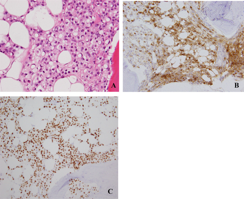
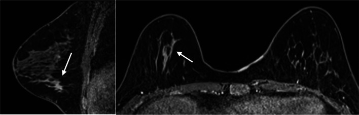
Case presentation: A 51-year-old woman presented to our outpatient clinic with dyspnea on exertion. She had severe anemia (hemoglobin, 5.3 g/dL) and thrombocytopenia (platelet count, 31 × 103/mL). Bone-marrow biopsy was performed to evaluate hematopoietic function. The pathologic diagnosis was bone-marrow carcinomatosis due to metastatic breast cancer. Initial mammography followed by ultrasonography (US) failed to detect the primary tumor. On MRI, a non-mass-enhancement lesion was observed. While second-look US also did not detect the lesion, it was clearly visualized with RVS. We were finally able to biopsy the breast lesion. The pathologic diagnosis was ILC positive for both estrogen receptor and progesterone receptor, with 1 + immunohistochemical staining for human epidermal growth factor receptor 2. This case of ILC was characterized by bone-marrow metastasis. Due to decreased cell adhesion, the risk of bone-marrow metastasis is higher in ILC than in invasive ductal carcinoma, the most prevalent type of breast cancer. Biopsy of the primary lesion, which was initially only detected with MRI, was successfully performed with clear visualization during RVS, which is based on the fusion of MRI and US images.
Conclusion: In this case report and literature review, we describe the unique clinical characteristics of ILC and a strategy for identifying primary lesions that are initially only visualized with MRI.
Keywords: Invasive lobular carcinoma; MRI; Non-mass-enhancement lesion; RVS.
© 2023. The Author(s).
Conflict of interest statement
The authors declare that they have no competing interests.
Figures


References
LinkOut - more resources
Full Text Sources
Research Materials
