

A method for accurate and reproducible specimen alignment for insertion tests of cochlear implant electrode arrays
- PMID: 37204650
- PMCID: PMC11582122
- DOI: 10.1007/s11548-023-02930-1
A method for accurate and reproducible specimen alignment for insertion tests of cochlear implant electrode arrays
Abstract
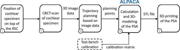
Purpose: The trajectory along which the cochlear implant electrode array is inserted influences the insertion forces and the probability for intracochlear trauma. Controlling the trajectory is especially relevant for reproducible conditions in electrode insertion tests. Using ex vivo cochlear specimens, manual alignment of the invisibly embedded cochlea is imprecise and hardly reproducible. The aim of this study was to develop a method for creating a 3D printable pose setting adapter to align a specimen along a desired trajectory toward an insertion axis.
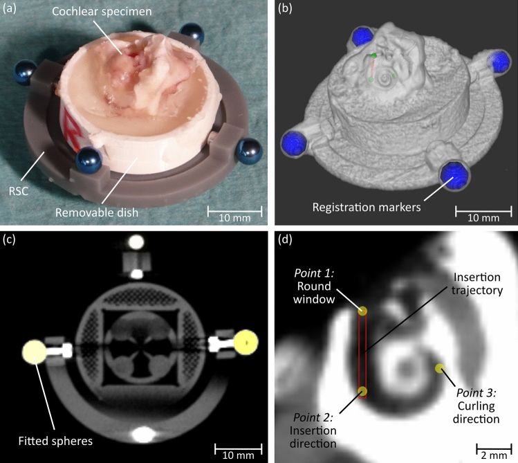
Methods: Planning points of the desired trajectory into the cochlea were set using CBCT images. A new custom-made algorithm processed these points for automated calculation of a pose setting adapter. Its shape ensures coaxial positioning of the planned trajectory to both the force sensor measuring direction and the insertion axis. The performance of the approach was evaluated by dissecting and aligning 15 porcine cochlear specimens of which four were subsequently used for automated electrode insertions.
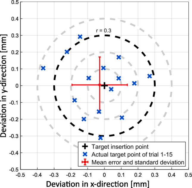
Results: The pose setting adapter could easily be integrated into an insertion force test setup. Its calculation and 3D printing was possible in all 15 cases. Compared to planning data, a mean positioning accuracy of 0.21 ± 0.10 mm at the level of the round window and a mean angular accuracy of 0.43° ± 0.21° were measured. After alignment, four specimens were used for electrode insertions, demonstrating the practical applicability of our method.
Conclusion: In this work, we present a new method, which enables automated calculation and creation of a ready-to-print pose setting adapter for alignment of cochlear specimens in insertion test setups. The approach is characterized by a high level of accuracy and reproducibility in controlling the insertion trajectory. Therefore, it enables a higher degree of standardization in force measurement when performing ex vivo insertion tests and thereby improves reliability in electrode testing.
Keywords: 3D printing; Electrode insertion tests; Porcine cochlea; Pose setting adapter; Specimen alignment.
© 2023. The Author(s).
Conflict of interest statement
The authors state that they have no conflict of interest.
Figures


References
-
- Gantz BJ, Turner C, Gfeller KE, Lowder MW (2005) Preservation of hearing in cochlear implant surgery: advantages of combined electrical and acoustical speech processing. Laryngoscope 115:796–802. 10.1097/01.MLG.0000157695.07536.D2 - PubMed
-
- Bas E, Dinh CT, Garnham C, Polak M, Van de Water TR (2012) Conservation of hearing and protection of hair cells in cochlear implant patients’ with residual hearing. Anat Rec 295:1909–1927. 10.1002/ar.22574 - PubMed
-
- Aebischer P, Mantokoudis G, Weder S, Anschuetz L, Caversaccio M, Wimmer W (2022) In-vitro study of speed and alignment angle in cochlear implant electrode array insertions. IEEE Trans Biomed Eng 69:129–137. 10.1109/TBME.2021.3088232 - PubMed
-
- Torres R, Drouillard M, De Seta D, Bensimon JL, Ferrary E, Sterkers O, Bernardeschi D, Nguyen Y (2018) Cochlear implant insertion axis into the basal turn: a critical factor in electrode array translocation. Otol Neurotol 39:168–176. 10.1097/MAO.0000000000001648 - PubMed
MeSH terms
Grants and funding
LinkOut - more resources
Full Text Sources
Medical
