. 2023 Apr 18;14(5):2040-2054.
doi: 10.1364/BOE.483835. eCollection 2023 May 1.

Signal attenuation-compensated projection-resolved OCT angiography

Affiliations

Signal attenuation-compensated projection-resolved OCT angiography

Jie Wang et al. Biomed Opt Express. .

Abstract

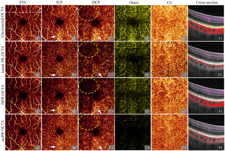
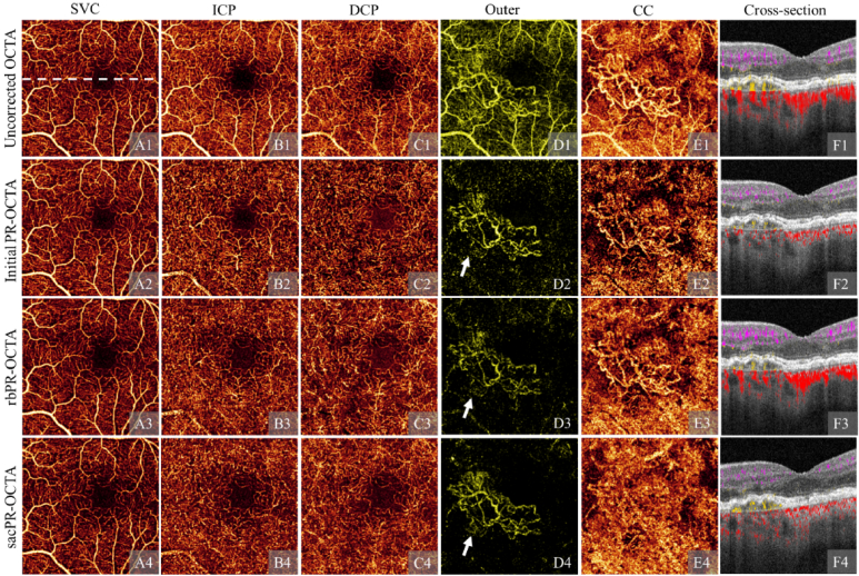
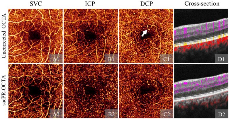
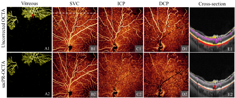
Projection artifacts are a significant limitation of optical coherence tomographic angiography (OCTA). Existing techniques to suppress these artifacts are sensitive to image quality, becoming less reliable on low-quality images. In this study, we propose a novel signal attenuation-compensated projection-resolved OCTA (sacPR-OCTA) algorithm. In addition to removing projection artifacts, our method compensates for shadows beneath large vessels. The proposed sacPR-OCTA algorithm improves vascular continuity, reduces the similarity of vascular patterns in different plexuses, and removes more residual artifacts compared to existing methods. In addition, the sacPR-OCTA algorithm better preserves flow signal in choroidal neovascular lesions and shadow-affected areas. Because sacPR-OCTA processes the data along normalized A-lines, it provides a general solution for removing projection artifacts agnostic to the platform.


Conflict of interest statement

Jie Wang: Optovue/Visionix, Inc (P, R); David Huang: Optovue/Visionix, Inc. (F, P, R), Boeringer Ingelheim Inc. (C); Yali Jia: Optovue/Visionix, Inc. (P, R), Optos Inc. (P).

Figures

Fig. 1.
Fig. 1.
Uncorrected OCTA with projection artifacts. (A) Cross-sectional OCT overlaid with color-coded OCTA signal (A) in the inner retina (violet), the outer retina (yellow), and the choroid (red) and the magnified view of the green box (A1). En face images are produced by maximum projection of the superficial vascular complex (SVC, B), intermediate capillaru plexus (ICP, C), deep capillary plexus (DCP, D), the outer retina (E), and the choriocapillaries (F) slabs.
Fig. 2.
Fig. 2.
Illustration of large vessel shadow compensation in structural OCT. (A) 3D structural OCT; (B) cross-section structural overlaid with a tissue mask (red) made using a 3D adaptive thresholding algorithm; (C) Cross-sectional structural OCT overlaid with the region of interested (ROI) mask (green) covering the slab of ganglion cell complex (corresponding to a 120 μm axial depth); (D) OCT angiogram generated in the superficial region using maximum projection. (E1) enhanced vasculature; (E2) Denoised OCT angiogram generated by multiplying the enhanced vasculature (E1) with the original OCT angiogram (D); (F) Large vessel mask detected by using adaptive thresholding algorithm. (G1) Original cross-sectional OCT with shadowing artifacts under the large vessels; (G2) Shadowing artifacts compensated cross-sectional OCT.
Fig. 3.
Fig. 3.
Signal attenuation-compensated projection resolved OCTA (sacPR-OCTA) in a healthy eye, showing the superficial vascular complex (SVC, A), the intermediate capillary plexus (ICP, B), the deep capillary plexus (DCP, C) and a cross-sectional structural OCT overlaid with the sacPR-OCTA color-coded according to anatomic location (violet: inner retina, yellow: outer retina red: choroid; position was highlighted by the dotted line in A). The results reflect the known features of retinal vascular anatomy from the histology [29].
Fig. 4.
Fig. 4.
A normal eye imaged with uncorrected OCTA (row 1), initial projection-resolved OCTA (PR-OCTA; row 2), reflectance-based projection-resolved OCTA (rbPR-OCTA, row 3), and the signal attenuation-compensated projection-resolved OCTA (sacPR-OCTA, row 4) showing en face OCT angiogram of the superficial vascular plexus (SVC, column A), the intermediate capillary plexus (ICP, column B), the deep capillary plexus (DCP, column C), the outer retina (column D) and the choriocapillaris (CC, column E), and the cross-sectional structural OCT overlaid with color-coded flow signal (column F; violet: inner retina, yellow: outer retina red: choroid) at the position indicated by the white dotted line in A1. The sacPR-OCTA removed more residual artifacts than the previous methods while maintaining vascular integrity, and volumetrically reduced the artifactual “tails” vessel tails (F1) with more anatomically correct circular shapes (F4) in cross-sectional OCTA. With both initial PR-OCTA and rbPR-OCTA, the flow signal is reduced under the larger vessels, particularly in the DCP and the CC (C2, E2, C3, E3), These overprocessing artifacts are absent in the proposed sacPR-OCTA method (C4, E4), which show uniform vascular patterns in the DCP and the CC.
Fig. 5.
Fig. 5.
A comparison of artifact suppression algorithms on a scan with shadow artifacts: uncorrected OCTA (row 1), initial projection-resolved OCTA (PR-OCTA, row 2), reflectance-based projection-resolved OCTA (rbPR-OCTA, row 3), and the signal attenuation-compensated projection-resolved OCTA (sacPR-OCTA, row 4) showing en face OCT angiogram of the superficial vascular plexus (SVC, column A), the intermediate capillary plexus (ICP, column B), the deep capillary plexus (DCP, column C), the outer retina (column D) and the choriocapillaris (CC, column E), and the cross-sectional structural OCT overlaid with color-coded flow signal (column F; violet: inner retina, yellow: outer retina red: choroid) The sacPR-OCTA algorithms preserved more of the true flow signal in the area affected by a shadow artifact (yellow dotted ellipses) and suppressed more projection artifacts (white arrows).
Fig. 6.
Fig. 6.
A comparison of artifact removal algorithms on a scan with choroidal neovascularization (CNV): uncorrected OCTA (row 1), initial projection-resolved OCTA (PR-OCTA, row 2), reflectance-based projection-resolved OCTA (rbPR-OCTA, row 3), and the signal attenuation-compensated projection-resolved OCTA (sacPR-OCTA, row 4) showing en face OCT angiogram of the superficial vascular plexus (SVC, column A), the intermediate capillary plexus (ICP, column B), the deep capillary plexus (DCP, column C), the outer retina (column D) and the choriocapillaris (CC, column E), and the cross-sectional structural OCT overlaid with color-coded flow signal (column F; violet: inner retina, yellow: outer retina red: choroid) The proposed sacPR-OCTA algorithm preserved more of the true CNV signal (highlighted by white arrows).
Fig. 7.
Fig. 7.
Signal attenuation-compensated projection-resolved OCTA (sacPR-OCTA) on angioplex 3 × 3-mm scan. Row 1-2: Uncorrected OCTA and sacPR-OCTA. Column A-C are en face angiograms of the superficial vascular plexus (SVC), intermediate capillary plexus (ICP), deep capillary plexus (DCP). Column D is cross-sectional structural OCT overlaid with color-coded flow signals (violet: inner retina, yellow: outer retina red: choroid). The white arrow highlights the dilated vessel in DCP. sacPR-OCTA removes projection artifacts in this widely-used commercial device.
Fig. 8.
Fig. 8.
Signal attenuation-compensated projection-resolved OCTA (sacPR-OCTA) on Solix 9 × 9-mm scan. Row 1-2: Uncorrected OCTA and sacPR-OCTA. Column A-D are en face angiograms of the neovascularization (NV) in the vitreous, superficial vascular plexus (SVC), intermediate capillary plexus (ICP), deep capillary plexus (DCP). Colum E is cross-sectional structural OCT overlaid with color-coded flow signals (gold: vitreous, violet: inner retina, yellow: outer retina red: choroid. The red arrow highlights the connection between the NV and feeder vessel. sacPR-OCTA removed projection artifacts in this wide field of view showing the optic nerve head in a newly released commercial device.

References

    1. Jia Y., Tan O., Tokayer J., Potsaid B., Wang Y., Liu J. J., Kraus M. F., Subhash H., Fujimoto J. G., Hornegger J., Huang D., “Split-spectrum amplitude-decorrelation angiography with optical coherence tomography,” Opt. Express 20(4), 4710–4725 (2012).10.1364/OE.20.004710 - DOI - PMC - PubMed
    1. An L., Wang R. K., “In vivo volumetric imaging of vascular perfusion within human retina and choroids with optical micro-angiography,” Opt. Express 16(15), 11438–11452 (2008).10.1364/OE.16.011438 - DOI - PubMed
    1. Hwang T. S., Gao S. S., Liu L., Lauer A. K., Bailey S. T., Flaxel C. J., Wilson D. J., Huang D., Jia Y., “Automated quantification of capillary nonperfusion using optical coherence tomography angiography in diabetic retinopathy,” JAMA Ophthalmol. 134(4), 367–373 (2016).10.1001/jamaophthalmol.2015.5658 - DOI - PMC - PubMed
    1. Zhang M., Hwang T. S., Dongye C., Wilson D. J., Huang D., Jia Y., “Automated quantification of nonperfusion in three retinal plexuses using projection-resolved optical coherence tomography angiography in diabetic retinopathy,” Invest. Ophthalmol. Visual Sci. 57, 5101–5106 (2016).10.1167/iovs.16-19776 - DOI - PMC - PubMed
    1. Nesper P. L., Soetikno B. T., Fawzi A. A., “Choriocapillaris nonperfusion is associated with poor visual acuity in eyes with reticular pseudodrusen,” Am. J. Ophthalmol. 174, 42–55 (2017).10.1016/j.ajo.2016.10.005 - DOI - PMC - PubMed

LinkOut - more resources