. 2023 Apr 12;14(5):1992-2002.
doi: 10.1364/BOE.488024. eCollection 2023 May 1.

Diffractive micro-lens array (DLA) for uniform and selective picosecond laser treatment

Affiliations

Diffractive micro-lens array (DLA) for uniform and selective picosecond laser treatment

Jongman Choi et al. Biomed Opt Express. .

Abstract

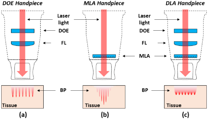
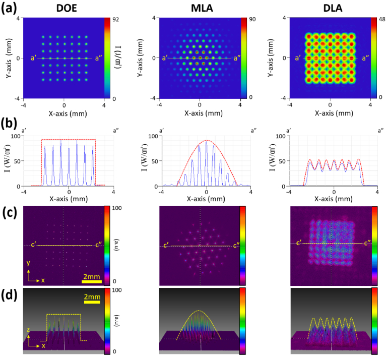
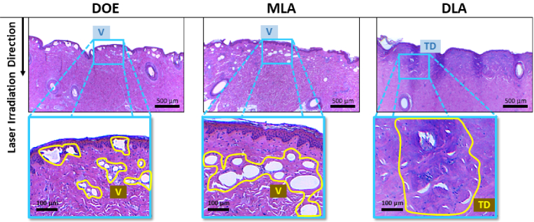
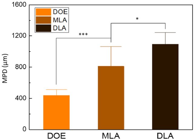
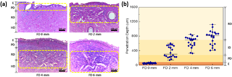
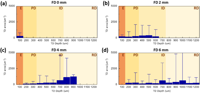
Picosecond Nd:YAG lasers using diffractive optical elements (DOE) and micro-lens arrays (MLA) have widely been used in dermatology for the treatment of pigmented lesions and skin rejuvenation. This study designed and developed a new optical element of diffractive micro-lens array (DLA) by combing the features of DOE and MLA in order to achieve uniform and selective laser treatment. Both optical simulation and beam profile measurement demonstrated that DLA created a square macro-beam consisting of multiple micro-beams in a uniform distribution. Histological analysis confirmed that the DLA-assisted laser treatment generated micro-injuries at various skin depths from the epidermal layer to the deep dermal layer (up to 1200 µm) by adjusting the focal depths while DOE showed shallow penetration depths and MLA created non-uniform micro-injury zones. The DLA-assisted picosecond Nd:YAG laser irradiation can provide a potential benefit for pigment removal and skin rejuvenation via uniform and selective laser treatment.


Conflict of interest statement

The authors declare that there are no conflicts of interest related to this article.

Figures

Fig. 1.
Fig. 1.
Schematic handpiece designs with three optical elements: (a) DOE, (b) MLA, and (c) DLA. Picosecond Nd:YAG laser light (red arrows) was perpendicularly incident on the skin tissue surface. Note that each tissue demonstrates the corresponding beam profile (DOE = diffractive optical elements; MLA = micro-lens array; DLA = diffractive micro-leans array; FL = focal lens; BP = beam profile).
Fig. 2.
Fig. 2.
Numerical simulations (a and b) and corresponding beam profiles (c and d) of DOE, MLA, and DLA: (a) simulated beam patterns at focal plane, (b) cross-sectional view of irradiance along line (á-á´) in (a), (c) measured beam profiles at focal plane, and (d) cross-sectional view of irradiance along line (ć-ć´) in (c).
Fig. 3.
Fig. 3.
Histological analysis of porcine skin tissue after picosecond Nd:YAG laser irradiation with DOE, MLA, and DLA: Picosecond Nd:YAG laser light was incident perpendicularly on the skin tissue surface with each optical element beam profile (red in top images; 40X). Note that the yellow solid lines in H&E-stained histological images (bottom; 200X) represent laser-induced vacuoles (V) by DOE and MLA) and thermal decomposition (TD) by DLA, respectively.
Fig. 4.
Fig. 4.
Quantitative comparison of maximum penetration depth (MPD in µm) in porcine skin tissue after picosecond laser treatment with DOE, MLA, and DLA (***p < 0.001 DOE vs. MLA; ***p < 0.05 DLA vs. DOE and MLA).
Fig. 5.
Fig. 5.
Quantitative analysis of porcine skin tissues after DLA-assisted laser irradiation at various focal depths (FD): (a) H&E-stained histological images (40X) captured at various FDs (0, 2, 4, and 6 mm). Yellow dotted areas represent laser-induced thermal decomposition (TD) in tissue. (b) Quantitative analysis on penetration depth of TD for each FD measured from histological images (N = 20). Note that different background colors in (b) represent various skin layers (E = epidermis; PD = papillary dermis; ID = intermediate region of the dermis; RD = reticular dermis).
Fig. 6.
Fig. 6.
Quantitative comparison of thermal decomposition (TD) area as function of TD penetration depth: (a) Focal depth (FD) = 0 mm, (b) FD = 2 mm, (c) FD = 4 mm, and (d) FD = 6 mm. Note that different background colors represent various skin layers (E = epidermis; PD = papillary dermis; ID = Intermediate region of the dermis; RD = reticular dermis).

References

    1. Dai Y. X., Chuang Y. Y., Chen P. Y., Chen C. C., “Efficacy and safety of ablative resurfacing with a high-energy 1,064 Nd-YAG picosecond-domain laser for the treatment of facial acne scars in Asians,” Lasers Surg. Med. 52(5), 389–395 (2020).10.1002/lsm.23151 - DOI - PubMed
    1. Kim D. G., Nam S. M., Shin J. S., Park E. S., “Effectiveness of the pico-toning technique for the treatment of melasma with a low fluence 1,064-nm Nd: YAG laser in Asian patients,” Medical Lasers 9, 166–171 (2020).10.25289/ML.2020.9.2.166 - DOI
    1. Carroll L., Humphreys T. R., “LASER-tissue interactions,” Clin. Dermatol. 24(1), 2–7 (2006).10.1016/j.clindermatol.2005.10.019 - DOI - PubMed
    1. Stewart N., Lim A. C., Lowe P. M., Goodman G., “Lasers and laser-like devices: Part one,” Australas. J. Dermatol. 54(3), 173–183 (2013).10.1111/ajd.12034 - DOI - PubMed
    1. Anderson R. R., Parrish J. A., “Selective photothermolysis: precise microsurgery by selective absorption of pulsed radiation,” Science 220(4596), 524–527 (1983).10.1126/science.6836297 - DOI - PubMed