. 2023 Apr 30;4(2):463-469.
doi: 10.1002/jha2.698. eCollection 2023 May.

Biallelic deleterious germline SH2B3 variants cause a novel syndrome of myeloproliferation and multi-organ autoimmunity

Affiliations

Biallelic deleterious germline SH2B3 variants cause a novel syndrome of myeloproliferation and multi-organ autoimmunity

Piers Blombery et al. EJHaem. .

Abstract

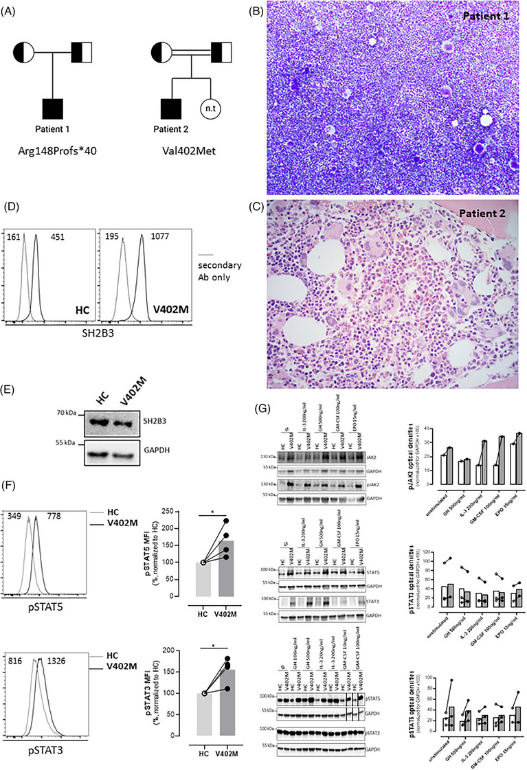
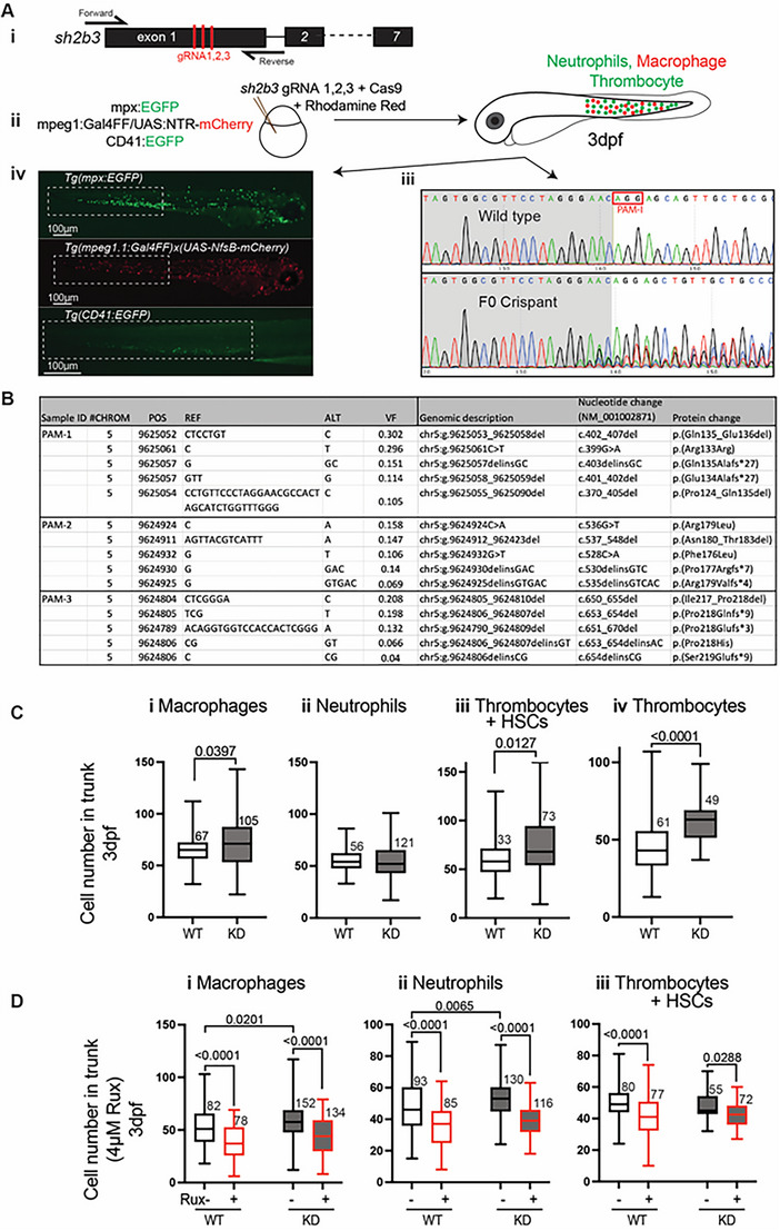
SH2B3 is a negative regulator of multiple cytokine receptor signalling pathways in haematopoietic tissue. To date, a single kindred has been described with germline biallelic loss-of-function SH2B3 variants characterized by early onset developmental delay, hepatosplenomegaly and autoimmune thyroiditis/hepatitis. Herein, we described two further unrelated kindreds with germline biallelic loss-of-function SH2B3 variants that show striking phenotypic similarity to each other as well as to the previous kindred of myeloproliferation and multi-organ autoimmunity. One proband also suffered severe thrombotic complications. CRISPR-Cas9 gene editing of zebrafish sh2b3 created assorted deleterious variants in F0 crispants, which manifest significantly increased number of macrophages and thrombocytes, partially replicating the human phenotype. Treatment of the sh2b3 crispant fish with ruxolitinib intercepted this myeloproliferative phenotype. Skin-derived fibroblasts from one patient demonstrated increased phosphorylation of JAK2 and STAT5 after stimulation with IL-3, GH, GM-CSF and EPO compared to healthy controls. In conclusion, these additional probands and functional data in combination with the previous kindred provide sufficient evidence for biallelic homozygous deleterious variants in SH2B3 to be considered a valid gene-disease association for a clinical syndrome of bone marrow myeloproliferation and multi-organ autoimmune manifestations.

Keywords: genetics; molecular diagnosis; myeloid function and development.


Conflict of interest statement

P.B. consulted for, advised, or received honoraria from Adaptive Biotechnologies, AstraZeneca, and Servier; S.B. has received personal fees or travel expenses from Immunodeficiency Canada/IAACI, CSL Behring, Baxalta US Inc., GSK and Biotest; G.J.L. has consulted for CSL Behring; J.L. has served on advisory boards for Kite/Gilead and received honoraria from Takeda.

Figures

FIGURE 1
FIGURE 1
Clinicopathological features of patients with biallelic loss of function variants in SH2B3. (A) Pedigree for patients 1 and 2; half‐filled squares and circles indicate carrier status for SH2B3 variants for males and females, respectively. Fully filled squares indicate homozygous SH2B3. n.t. – not tested. Double line indicates consanguinity. (B) Bone marrow aspirate from patient 1 demonstrating hypercellular marrow with megakaryocytic hyperplasia and morphological atypia (×100). (C) Bone marrow trephine biopsy from patient 2 demonstrated megakaryocytic hyperplasia and atypia (haematoxylin and eosin (H&E), ×400). (D) SH2B3 expression assessed intracellularly by flow cytometry on skin‐derived fibroblasts using a mouse anti‐human SH2B3 antibody followed by goat anti‐mouse IgG‐AlexaFluor488 staining after fixation and permeabilization using the BD Cytofix/CytoPerm buffers. Immunoreactive SH2B3 expression is present in wildtype healthy control (HC) and proband (V402M) cells. Negative control is secondary antibody (Ab) only. Numbers inside panels represent mean fluorescence intensity (MFI). (E) Western blot showing normal expression of SH2B3 in V402M skin‐derived fibroblasts; GADPH loading control. (F) Basal STAT‐5 and STAT‐3 phosphorylation was evaluated on skin‐derived fibroblasts by Phosflow assay using BD Phosflow Fix I and Perm III buffers according to manufacturer instructions (BD Biosciences). Histograms on the left show a representative example and graphs on the right the normalised mean values for four experiments. Numbers inside histograms represent mean fluorescence intensity (MFI). pSTAT3 and pSTAT5 levels were significantly elevated relative to control in the proband (V402M) samples (*indicates p < 0.05, Mann–Whitney test). (G) Increased phosphorylation of JAK2 and STAT5 in SH2B3‐V402M. Skin‐derived fibroblasts were obtained from patient 2 and from a healthy control were assessed for SH2B3, pJAK2, pSTAT3 and pSTAT5 protein expression by immunoblotting before and after 15‐min stimulation with the indicated ligands (optical density for experiment and replicates provided in histogram). Black lines next to samples in lanes 13 and 14 (pSTAT5 and GAPDH) indicate where images have been moved in the figure to allow comparison between healthy control versus patient. Graphs on the right show quantitation of multiple immunoblots with lines connecting paired samples. EPO, erythropoietin; GH, growth hormone; GM‐CSF, granulocyte‐macrophage colony‐stimulating factor; HC, healthy control; IL‐3, interleukin‐3.
FIGURE 2
FIGURE 2
Zebrafish sh2b3 loss‐of‐function model. (Ai) CRISPR/Cas9‐mediated mutagenesis to truncate the sh2b3 at exon 1. Red bars indicate the target sites of three designed gRNAs. (Aii) Microinjection of 3x gRNAs into one‐cell stage zebrafish embryo along with Cas9 protein and rhodamine. (Aiii) Sanger sequencing of sh2b3 locus in wild type (top) and F0 crispant (bottom) zebrafish embryos indicating on‐target gene disruption at gRNA‐1 target site (as an example). The red rectangle indicates the PAM sequence for the gRNA‐1. (Aiv) Respectively, EGFP and mCherry expressing neutrophils and macrophages in Tg(mpx:EGFP) and Tg(mpeg1.1:Gal4FF/UAS:NfsB‐mCherry), and EGFP expressing cells (HSCs and thrombocytes) in Tg (CD41:EGFP) 3dpf zebrafish embryos. Dashed white rectangles mark regions in which cells have been quantified. (B) NGS result showing the top 5 most common variants at each gRNA target site. (C) Quantification of macrophages (mpeg1:mCherry positive), neutrophils (mpx:EGFP positive), HSCs and thrombocytes (CD41:EGFP positive) and thrombocytes (mpl:EGFP positive) in the tail region of 3 dpf sh2b3 knockdown (F0 crispants) compared to wildtype embryos. (D) Quantification of macrophages, neutrophils and CD41:EGFP positive cells in sh2b3 knockdown and wildtype embryos in the presence or absence of 4 μM ruxolitinib. HSCs, hematopoietic stem cells; gRNA, guide RNA; dpf, days post fertilization; KD, knockdown; PAM, protospacer adjacent motif; VF, variant frequency; Rux, Ruxolitinib; WT, wild‐type. Box and whisker plots (range, 25th and 75th percentile, median) for n embryos (n values near box) pooled from three biologically independent experiments. p‐Values from unpaired T‐test (C) and one‐way ANOVA with Tukey's multiple comparison test (D); p‐values shown only for significant differences.

References

    1. Tong W, Zhang J, Lodish HF. Lnk inhibits erythropoiesis and Epo‐dependent JAK2 activation and downstream signaling pathways. Blood. 2005;105(12):4604–12. - PMC - PubMed
    1. Gery S, Cao Q, Gueller S, Xing H, Tefferi A, Koeffler HP. Lnk inhibits myeloproliferative disorder‐associated JAK2 mutant, JAK2V617F. J Leukoc Biol. 2009;85(6):957–65. - PMC - PubMed
    1. Hurtado C, Erquiaga I, Aranaz P, Miguéliz I, García‐Delgado M, Novo FJ, et al. LNK can also be mutated outside PH and SH2 domains in myeloproliferative neoplasms with and without V617FJAK2 mutation. Leuk Res. 2011;35(11):1537–9. - PubMed
    1. Maslah N, Cassinat B, Verger E, Kiladjian JJ, Velazquez L. The role of LNK/SH2B3 genetic alterations in myeloproliferative neoplasms and other hematological disorders. Leukemia. 2017;31(8):1661–70. - PubMed
    1. Perez‐Garcia A, Ambesi‐Impiombato A, Hadler M, Rigo I, LeDuc CA, Kelly K, et al. Genetic loss of SH2B3 in acute lymphoblastic leukemia. Blood. 2013;122(14):2425–32. - PMC - PubMed

LinkOut - more resources