

Anthocyanins from Malus spp. inhibit the activity of Gymnosporangium yamadae by downregulating the expression of WSC, RLM1, and PMA1
- PMID: 37206329
- PMCID: PMC10191115
- DOI: 10.3389/fmicb.2023.1152050
Anthocyanins from Malus spp. inhibit the activity of Gymnosporangium yamadae by downregulating the expression of WSC, RLM1, and PMA1
Abstract
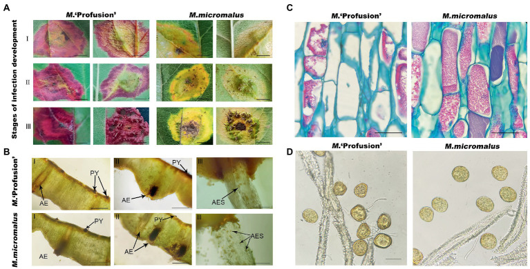
Malus plants are frequently devastated by the apple rust caused by Gymnosporangium yamadae Miyabe. When rust occurs, most Malus spp. and cultivars produce yellow spots, which are more severe, whereas a few cultivars accumulate anthocyanins around rust spots, forming red spots that inhibit the expansion of the affected area and might confer rust resistance. Inoculation experiments showed that Malus spp. with red spots had a significantly lower rust severity. Compared with M. micromalus, M. 'Profusion', with red spots, accumulated more anthocyanins. Anthocyanins exhibited concentration-dependent antifungal activity against G. yamadae by inhibiting teliospores germination. Morphological observations and the leakage of teliospores intracellular contents evidenced that anthocyanins destroyed cell integrity. Transcriptome data of anthocyanins-treated teliospores showed that differentially expressed genes were enriched in cell wall and membrane metabolism-related pathways. Obvious cell atrophy in periodical cells and aeciospores was observed at the rust spots of M. 'Profusion'. Moreover, WSC, RLM1, and PMA1 in the cell wall and membrane metabolic pathways were progressively downregulated with increasing anthocyanins content, both in the in vitro treatment and in Malus spp. Our results suggest that anthocyanins play an anti-rust role by downregulating the expression of WSC, RLM1, and PMA1 to destroy the cell integrity of G. yamadae.
Keywords: Gymnosporangium yamadae; anthocyanins; cell membrane; cell wall; teliospores; transcriptome.
Copyright © 2023 Wang, An, Guo, Wang, Shang, Chen, Liu, Meng, Zhang, Wei and Li.
Conflict of interest statement
The author declares that the research was conducted in the absence of any commercial or financial relationships that could be construed as a potential conflict of interest.
Figures


References
-
- Bautista-Silva J. P., Seibert J. B., Amparo T. R., Rodrigues I. V., Teixeira L. F. M., Souza G. H. B., et al. . (2020). Melaleuca leucadendra essential oil promotes loss of cell membrane and wall integrity and inhibits bacterial growth: an in silico and in vitro approach. Curr. Microbiol. 77, 2181–2191. doi: 10.1007/s00284-020-02024-0, PMID: - DOI - PubMed
-
- Chen Y. (2014). Functional characterization of 20 signaling and effector proteins essential for cell wall integrity and their connections to biocontrol potential of entomopathogenic fungi. PhD thesis, Hangzhou, Zhejiang, China: Zhejiang University.
-
- Deng H. T., Zhu J. Y., Tong Y. Q., Kong Y. W., Tan C., Wang M. Y., et al. . (2021). Antibacterial characteristics and mechanisms of action of Aronia melanocarpa anthocyanins against Escherichia coli. Lwt-Food Sci. Technol. 150:112018. doi: 10.1016/j.lwt.2021.112018 - DOI
-
- Duan Y. D., Hao S. X., Luo R., Lu Y. F., Li G., Zhang J., et al. . (2019). Antioxidant defense against rust infection in the leaf tissue of Malus crabapple. Acta Physiol. Plant. 41:13. doi: 10.1007/s11738-019-2849-2 - DOI
LinkOut - more resources
Full Text Sources
Miscellaneous