Review
. 2023 May 2;14(19):4961-4978.
doi: 10.1039/d3sc00439b. eCollection 2023 May 17.

Aptamers 101: aptamer discovery and in vitro applications in biosensors and separations

Affiliations
Review

Aptamers 101: aptamer discovery and in vitro applications in biosensors and separations

Lucy F Yang et al. Chem Sci. .

Abstract

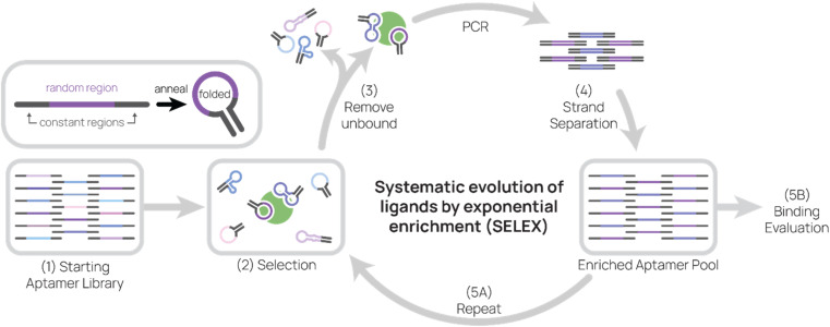
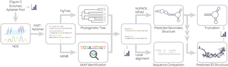
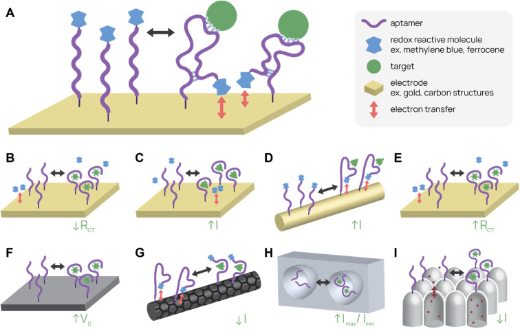
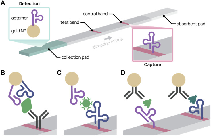
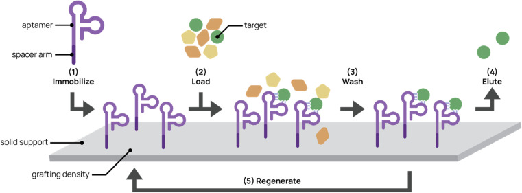
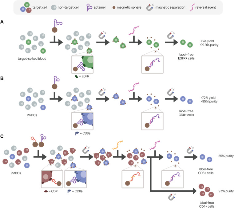
Aptamers are single-stranded nucleic acids that bind and recognize targets much like antibodies. Recently, aptamers have garnered increased interest due to their unique properties, including inexpensive production, simple chemical modification, and long-term stability. At the same time, aptamers possess similar binding affinity and specificity as their protein counterpart. In this review, we discuss the aptamer discovery process as well as aptamer applications to biosensors and separations. In the discovery section, we describe the major steps of the library selection process for aptamers, called systematic evolution of ligands by exponential enrichment (SELEX). We highlight common approaches and emerging strategies in SELEX, from starting library selection to aptamer-target binding characterization. In the applications section, we first evaluate recently developed aptamer biosensors for SARS-CoV-2 virus detection, including electrochemical aptamer-based sensors and lateral flow assays. Then we discuss aptamer-based separations for partitioning different molecules or cell types, especially for purifying T cell subsets for therapeutic applications. Overall, aptamers are promising biomolecular tools and the aptamer field is primed for expansion in biosensing and cell separation.


Conflict of interest statement

There are no conflicts to declare.

Figures

Fig. 1
Fig. 1. Systematic evolution of ligands by exponential enrichment (SELEX). Aptamers are composed of a random region and constant regions. After annealing, aptamers fold and adopt secondary structures that may allow them to bind to targets. (1) The starting aptamer library contains 1014–1015 unique sequences. (2) Then the aptamer library is incubated with target during selection. (3) Unbound aptamers are removed, and target-binding aptamers are eluted. (4) After conducting PCR on aptamers, sense and anti-sense strands are separated. (5) The enriched aptamer pool is used in the next round of selection and evaluated for binding.
Fig. 2
Fig. 2. Representative sequence analysis pipeline. The enriched aptamer pool(s) from SELEX were sequenced, and the data set was analyzed to produce a list of aptamer sequences and their relative abundance. This list was used to generate a phylogenetic tree and to search for motifs. Combining information from these analyses, candidate aptamers were chosen for further investigation. The predicted 2D structure and sequence similarity to other aptamers were generated based on the candidate aptamer sequence. From the predicted 2D structure, truncations were designed and 3D structure were predicted. At several points (bar graph icon), binding studies were conducted.
Fig. 3
Fig. 3. Electrochemical aptamer-based (E-AB) biosensor diagrams. (A) In a typical “signal-on” E-AB, aptamers change conformation after target binding, resulting in an increase in electrical signal. (B–I) SARS-CoV-2 detecting E-ABs. (B) Virus detection from Lasserre et al. (C) Spike protein detection from Martínez-Roque et al. (D) Spike protein detection from Idili et al. (E) Virus detection by “Cov-eChip” from Zhang et al. (F) Virus detection from Ban et al. (G) Spike protein detection from Curti et al. (H) Virus detection from Peinetti et al. (I) Virus detection from Shi et al.
Fig. 4
Fig. 4. Aptamer lateral flow assay (LFA) diagrams. (A) In a typical aptamer sandwich LFA, target molecules within a sample bind both the aptamer-conjugated gold nanoparticle (NP) detection agent (green) and the aptamer capture agent (pink), turning the test band dark. (B)–(D) Schematics showing only the test bands during detection. (B) Aptamer-antibody LFA detecting SARS-CoV-2 nucleocapsid protein (green) with two different aptamer sequences on the gold NP. Adapted from Zhang et al. (C) Antibody-free aptamer LFA (“AptaFlow”) detecting SARS-CoV-2 virus (green) with one aptamer for capture and another for detection. Adapted from Yang et al. (D) Multiplex LFA for detecting two variants of SARS-CoV-2 spike protein (green and teal) using two test bands with unique aptamers. Adapted from Yang et al.
Fig. 5
Fig. 5. Aptamers for affinity chromatography. (1) Aptamers designed with spacer arms are immobilized onto a solid support. (2) Mixed input is loaded onto the platform and aptamer captures the target. (3) The platform is washed to remove non-targets. (4) The target is eluted from the support. (5) In some cases, the platform can be regenerated for repeated use.
Fig. 6
Fig. 6. Aptamer-based platforms for label-free cell isolation. A mixed cell population is incubated with aptamer-labeled magnetic spheres, which bind specifically to the target cell. After magnetic separation to remove non-target cells (gray), labeled target cells remain. A reversal agent, or oligonucleotide complementary to a sequence within the aptamer, is added to release aptamer binding, resulting in a label-free target cell population. (A) Separation of EGFR+ cells (green) spiked in blood. Adapted from Gray et al. (B) Separation of CD8+ cells (purple) from PMBCs. Adapted from Kacherovsky et al. (C) Serial elution of CD4+ (red) and CD8+ (purple) cells from PBMCs. Adapted Cheng et al.
None
Lucy F. Yang
None
Melissa Ling
None
Nataly Kacherovsky
None
Suzie H. Pun

References

    1. Alves Ferreira-Bravo I. Cozens C. Holliger P. DeStefano J. J. Nucleic Acids Res. 2015;43:9587–9599. - PMC - PubMed
    1. Chen L. Rashid F. Shah A. Awan H. M. Wu M. Liu A. Wang J. Zhu T. Luo Z. Shan G. Proc. Natl. Acad. Sci. U. S. A. 2015;112:10002–10007. doi: 10.1073/pnas.1502159112. - DOI - PMC - PubMed
    1. Zhou J. Rossi J. Nat. Rev. Drug Discovery. 2017;16:181–202. doi: 10.1038/nrd.2016.199. - DOI - PMC - PubMed
    1. Liu J. Lu Y. Nat. Protoc. 2006;1:246–252. doi: 10.1038/nprot.2006.38. - DOI - PubMed
    1. Kosuri S. Church G. M. Nat. Methods. 2014;11:499–507. doi: 10.1038/nmeth.2918. - DOI - PMC - PubMed