. 2023 Apr 26;25(6):282.
doi: 10.3892/etm.2023.11981. eCollection 2023 Jun.

Expression of S100A9 in adamantinomatous craniopharyngioma and its association with wet keratin formation

Affiliations

Expression of S100A9 in adamantinomatous craniopharyngioma and its association with wet keratin formation

Chuan Zhao et al. Exp Ther Med. .

Abstract

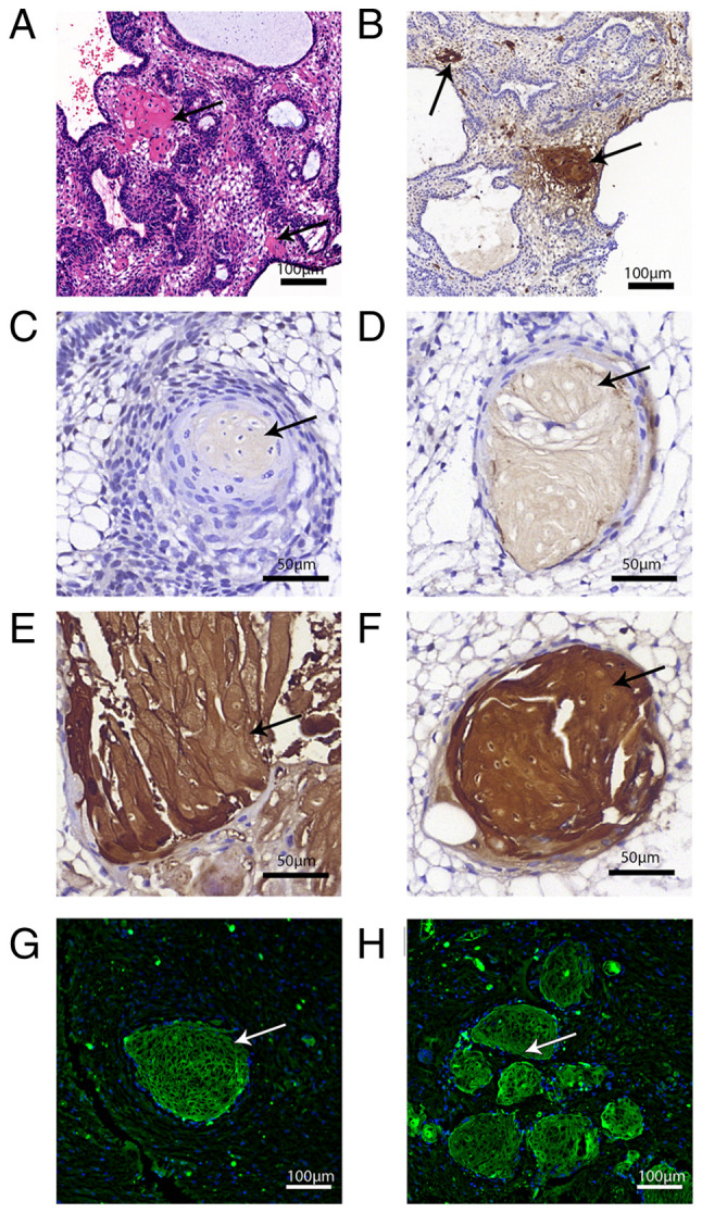
Wet keratin is a hallmark of adamantinomatous craniopharyngioma (ACP), which is frequently infiltrated by inflammatory cells. S100 calcium-binding protein A9 (S100A9) has been confirmed to play a decisive role in the development of inflammation. However, the relationship between wet keratin (keratin nodules) and S100A9 in ACP is poorly understood. The objective of the present study was to explore the expression of S100A9 in ACP and its association with wet keratin formation. Immunohistochemistry and immunofluorescence were used to detect the expression of S100A9, β-catenin and Ki67 in 46 cases of ACP. A total of three online databases were used to analyze S100A9 gene expression and protein data. The results revealed that S100A9 was primarily expressed in wet keratin and some intratumoral and peritumoral cells, and its expression in wet keratin was upregulated in the high inflammation group (P=1.800x10⁻³). In addition, S100A9 was correlated with the degree of inflammation (r=0.6; P=7.412x10⁻³) and the percentage of Ki67-positive cells (r=0.37; P=1.000x10⁻²). In addition, a significant correlation was noted between the area of wet keratin and the degree of inflammation (r=0.51; P=2.500x10⁻⁴). In conclusion, the present study showed that S100A9 was upregulated in ACP and may be closely associated with wet keratin formation and the infiltration of inflammatory cells in ACP.

Keywords: Ki67; S100 calcium-binding protein A9; adamantinomatous craniopharyngioma; inflammation; wet keratin.


Conflict of interest statement

The authors declare that they have no competing interests.

Figures

Figure 1
Figure 1
Online databases validate the expression of S100A9 in craniopharyngioma. (A) mRNA expression of S100A9 in pediatric brain tumors. (B) Protein expression of S100A9 in pediatric brain tumors. (C) The S100A9 gene located on chromosome 1q21.3 involved in the formation of epidermal differentiation complex had complex protein-protein interactions with epidermis-related SPRR genes and S100A family genes. S100A9, S100 calcium-binding protein A9; SPRR, small proline-rich protein.
Figure 2
Figure 2
Stepwise evolution of wet keratin in ACP. (A) The pathological structure of ACP. (B) Inflammatory cells were extensively aggregated in the ACP high-inflammation group (arrow). (C) In the ACP low-inflammation group, distribution of inflammatory cells was scattered (arrow). (D) The wet keratin-like tumor cells gradually formed wet keratin and their nuclei gradually became smaller (arrow). (E) Wet keratin was formed, the nuclei disappeared and a cavity was then formed (arrow). (F) The wet keratin content increased, and layered and fused together to form a flaky area (arrow). (G) Clear blue calcium salt deposits appeared on wet keratin (arrow). (H) Wet keratin appeared in the invasive margin of the tumor and in the surrounding brain tissue and formed an isolated island shape (arrow). (I) Multiple wet keratins infiltrated into the surrounding brain tissue. ACP, adamantinomatous craniopharyngioma; wk, wet keratin; cluster, cells with nucleocytoplasmic accumulation of β-catenin; SR, stellate reticulum; PE, palisade-like basal epithelium; GBT, gliotic brain tissue.
Figure 3
Figure 3
S100A9 is expressed in ACP. (A) Immunohistochemistry of β-catenin on ACP. (B) β-catenin staining for wet keratin was negative. (C) The cell cluster had nucleocytoplasmic accumulation of β-catenin. Fig. 3A-C are derived from the same sample. (D) No obvious dark brown staining of wet keratin was found in the negative control images. (E) S100A9 expression was positive in wet keratin (arrow; x5 objective). (F) S100A9 was expressed in the stroma surrounding the tumor (arrow). (G) S100A9 was partially expressed in stellate reticulum (arrow). (H) S100A9 showed weak positive staining in whorl-like cells in some cases (arrow). (I) S100A9 expression was positive in wet keratin (arrow; x20 objective) (J) Immunofluorescence staining showed that S100A9 was expressed on ACP tumor cells (green; red arrow). (K) β-catenin staining was nuclear positive in ACP cell cluster (red; arrow). (L) The merge of S100A9 and β-catenin showed that S100A9 was expressed around and inside whorl-like tumor cells (green; red arrow). ACP, adamantinomatous craniopharyngioma; S100A9, S100 calcium-binding protein A9.
Figure 4
Figure 4
Wet keratin-like cells in H&E staining and wet keratin in immunostaining. (A) Wet keratin-like cells in H&E-stained tumor cells with eosinophilic features (arrow). (B) Patchy S100A9-positive areas in tumor cells, corresponding to H&E-stained wet keratin-like tumor cells (arrow). (C) The transitional morphology of tumor cells to wet keratin; the nucleus shrank and the cytolysis expanded (arrow). (D) Wet keratin was further formed, the nucleus shrank further into dark brown spots, and S100A9 staining intensified (arrow). (E) The previously shrunken nuclei disappeared, and the wet keratin area was larger, with some modules layered in long strips and fused with each other (arrow). (F) Circular stained area of wet keratin. (G) Immunofluorescence staining showed that a single S100A9-positive wet keratin infiltrated into the brain tissue surrounding the tumor (arrow). (H) Multiple S100A9-positive wet keratins infiltrated into the brain tissue surrounding the tumor (arrow). H&E, hematoxylin and eosin; S100A9, S100 calcium-binding protein A9.
Figure 5
Figure 5
Wet keratin, S100A9 and inflammation. (A) H&E staining showed inflammatory cells (arrows) in the brain tissue surrounding the tumor margin. (B) Immunohistochemistry showed S100A9-positive areas (arrow) in brain tissue adjacent to the tumor. (C) H&E staining showed cholesterol clefts (arrowhead) surrounded by massive inflammatory cell infiltration (arrow). (D) Immunohistochemistry showed cholesterol clefts (arrowhead) surrounding evident S100A9 expression. (E) No or weak expression of S100A9 was observed in the intratumor and stroma of tumors in the low-inflammatory tumor group. (F) Strong expression of S100A9 was observed in both intratumor and stroma of tumors in the high-inflammatory tumor group. (G) The area of wet keratin was positively correlated with ACP inflammation (P=2.500x10⁻⁴). (H) S100A9 expression in wet keratin was positively correlated with inflammation in ACP (P=7.412x10⁻³). (I) Comparison of the S100A9 expression scores in the high-inflammation group and the low-inflammation group, with significantly higher S100A9 scores in the high inflammation group (P=1.800x10⁻³, t-test). S100A9, S100 calcium-binding protein A9; H&E, hematoxylin and eosin; ACP, adamantinomatous craniopharyngioma.
Figure 6
Figure 6
S100A9, Ki67 and prognosis. (A) Immunohistochemical staining for Ki67 showed no positive cells in the wet keratin area, but positive cells were found in scattered areas within the tumor and around the wet keratin (arrow, wet keratin; arrowhead, Ki67). (B) Immunofluorescence co-staining of S100A9 and Ki67. (C) A clear linear relationship was observed between S100A9 scores and the percentage of Ki67-positive cells (P=1.000x10⁻²). (D) S100A9 showed a trend of influencing recurrence-free survival of patients (P=6.200x10⁻²). S100A9, S100 calcium-binding protein A9.

References

    1. Müller H, Merchant T, Warmuth-Metz M, Martinez-Barbera J, Puget S. Craniopharyngioma. Nat Rev Dis Primers. 2019;5(75) doi: 10.1038/s41572-019-0125-9. - DOI - PubMed
    1. Louis DN, Perry A, Reifenberger G, von Deimling A, Figarella-Branger D, Cavenee WK, Ohgaki H, Wiestler OD, Kleihues P, Ellison DW. The 2016 World Health Organization classification of tumors of the central nervous system: A summary. Acta Neuropathol. 2016;131:803–820. doi: 10.1007/s00401-016-1545-1. - DOI - PubMed
    1. Martinez-Barbera J. Molecular and cellular pathogenesis of adamantinomatous craniopharyngioma. Neuropathol Appl Neurobiol. 2015;41:721–732. doi: 10.1111/nan.12226. - DOI - PMC - PubMed
    1. Whelan R, Prince E, Gilani A, Hankinson T. The inflammatory milieu of adamantinomatous craniopharyngioma and its implications for treatment. J Clin Med. 2020;9(519) doi: 10.3390/jcm9020519. - DOI - PMC - PubMed
    1. Wang S, Song R, Wang Z, Jing Z, Wang S, Ma J. S100A8/A9 in inflammation. Front Immunol. 2018;9(1298) doi: 10.3389/fimmu.2018.01298. - DOI - PMC - PubMed