

A Large Facial Nerve Schwannoma Presenting as a Swelling in Parotid Region-A Rare Case Report
- PMID: 37206751
- PMCID: PMC10188757
- DOI: 10.1007/s12070-022-03423-4
A Large Facial Nerve Schwannoma Presenting as a Swelling in Parotid Region-A Rare Case Report
Abstract
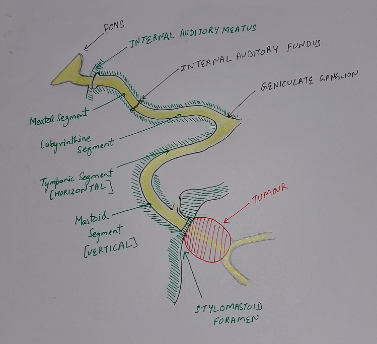
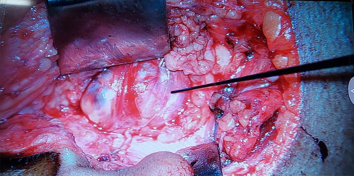
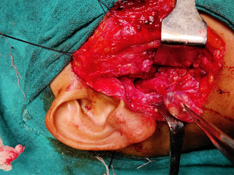
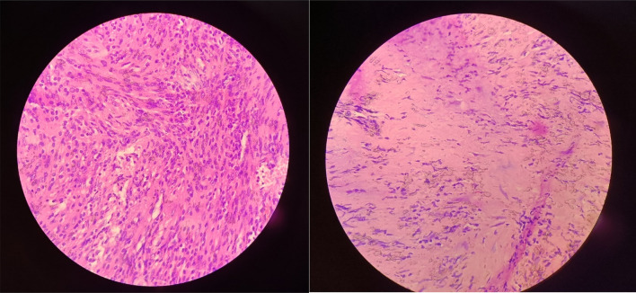
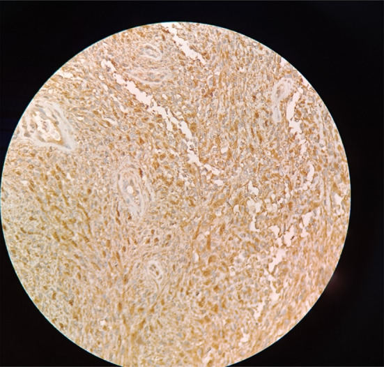
Extratemporal facial nerve schwannomas are rare entity. Pre-operative assessment is mostly inconclusive with differential diagnosis of parotid tumors. We hereby report a case of 28 years female who presented with painless swelling in right parotid region with normal facial nerve function. Ultrasonography was suggestive well circumscribed and homogenous mass arising from the deep lobe of parotid gland. The Fine-needle aspiration cytology came out to be inconclusive. For further characterization of the tumor contrast enhanced MR imaging was performed. The MR imaging revealed well-defined cystic, heterogeneous, pear-shaped mass lesion situated near the stylomastoid foramen. Post operatively the mass came out to be schwannoma on histopathological examination.
Keywords: Extratemporal; Facial nerve; Histopathological examination; Schwannoma.
© Association of Otolaryngologists of India 2023, Springer Nature or its licensor (e.g. a society or other partner) holds exclusive rights to this article under a publishing agreement with the author(s) or other rightsholder(s); author self-archiving of the accepted manuscript version of this article is solely governed by the terms of such publishing agreement and applicable law.
Conflict of interest statement
Conflict of interestAll author declares no conflict of interest.
Figures


References
LinkOut - more resources
Full Text Sources