Frozen Elephant Trunk Technique to Treat Extensive Thoracic Aortic Mural Thrombus
- PMID: 37207013
- PMCID: PMC10191685
- DOI: 10.1055/s-0041-1740916
Frozen Elephant Trunk Technique to Treat Extensive Thoracic Aortic Mural Thrombus
Abstract
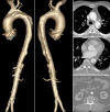
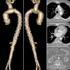
We describe a case report of a 63-year-old man who presented with chronic left-hand weakness and the absence of a pulse in the left arm. Thoracoabdominal computed tomography (CT) revealed an extensive thoracic aortic mural thrombus. Initial anticoagulation therapy did not provide a positive result, so the patient was referred for surgery. Hybrid aortic arch surgery using the frozen elephant trunk technique was performed with excellent early outcomes. A CT performed in the early postoperative period showed that the thrombus was completely excluded from the aortic lumen by the hybrid graft. No thrombus dislodgment was detected. No thrombus recurrence was observed during 19 months of follow-up.
Keywords: aortic thrombus; frozen elephant trunk; thoracic aorta.
International College of Angiology. This article is published by Thieme.
Conflict of interest statement
Conflict of Interest The authors declare no conflicts of interest.
Figures


Similar articles
-
Reversed Frozen Elephant Trunk Technique to Treat a Type II Thoracoabdominal Aortic Aneurysm.J Endovasc Ther. 2017 Apr;24(2):277-280. doi: 10.1177/1526602816678992. Epub 2016 Nov 15. J Endovasc Ther. 2017. PMID: 28112018
-
Fate of distal aorta after frozen elephant trunk and total arch replacement for type A aortic dissection in Marfan syndrome.J Thorac Cardiovasc Surg. 2019 Mar;157(3):835-849. doi: 10.1016/j.jtcvs.2018.07.096. Epub 2018 Aug 24. J Thorac Cardiovasc Surg. 2019. PMID: 30635189
-
Bail-out thoracic endovascular aortic repair for incorrect deployment of frozen elephant trunk into the false lumen.Interact Cardiovasc Thorac Surg. 2020 Jun 1;30(6):947-949. doi: 10.1093/icvts/ivaa043. Interact Cardiovasc Thorac Surg. 2020. PMID: 32236537
-
Downstream thoracic endovascular aortic repair following the frozen elephant trunk procedure.Cardiovasc Diagn Ther. 2022 Jun;12(3):272-277. doi: 10.21037/cdt-22-99. Cardiovasc Diagn Ther. 2022. PMID: 35800359 Free PMC article. Review.
-
The frozen elephant trunk procedure: indications, outcomes and future directions.Cardiovasc Diagn Ther. 2022 Oct;12(5):708-721. doi: 10.21037/cdt-22-330. Cardiovasc Diagn Ther. 2022. PMID: 36329958 Free PMC article. Review.
References
-
- Verma H, Meda N, Vora S, George R K, Tripathi R K. Contemporary management of symptomatic primary aortic mural thrombus. J Vasc Surg. 2014;60(06):1524–1534. - PubMed
-
- Rancic Z, Pfammatter T, Lachat M, Frauenfelder T, Veith F J, Mayer D. Floating aortic arch thrombus involving the supraaortic trunks: successful treatment with supra-aortic debranching and antegrade endograft implantation. J Vasc Surg. 2009;50(05):1177–1180. - PubMed
-
- Tsilimparis N, Hanack U, Pisimisis G, Yousefi S, Wintzer C, Rückert R I. Thrombus in the non-aneurysmal, non-atherosclerotic descending thoracic aorta–an unusual source of arterial embolism. Eur J Vasc Endovasc Surg. 2011;41(04):450–457. - PubMed

