. 2023 Jun 23;51(11):5341-5350.
doi: 10.1093/nar/gkad434.

Dynamic accumulation of cyclobutane pyrimidine dimers and its response to changes in DNA conformation

Affiliations

Dynamic accumulation of cyclobutane pyrimidine dimers and its response to changes in DNA conformation

Ravina Moirangthem et al. Nucleic Acids Res. .

Abstract

Photochemical dimerization of adjacent pyrimidines is fundamental to the creation of mutagenic hotspots caused by ultraviolet light. Distribution of the resulting lesions (cyclobutane pyrimidine dimers, CPDs) is already known to be highly variable in cells, and in vitro models have implicated DNA conformation as a major basis for this observation. Past efforts have primarily focused on mechanisms that influence CPD formation and have rarely considered contributions of CPD reversion. However, reversion is competitive under the standard conditions of 254 nm irradiation as illustrated in this report based on the dynamic response of CPDs to changes in DNA conformation. A periodic profile of CPDs was recreated in DNA held in a bent conformation by λ repressor. After linearization of this DNA, the CPD profile relaxed to its characteristic uniform distribution over a similar time of irradiation to that required to generate the initial profile. Similarly, when a T tract was released from a bent conformation, its CPD profile converted under further irradiation to that consistent with a linear T tract. This interconversion of CPDs indicates that both its formation and reversion exert control on CPD populations long before photo-steady-state conditions are achieved and suggests that the dominant sites of CPDs will evolve as DNA conformation changes in response to natural cellular processes.


Figures

Graphical Abstract
Graphical Abstract
Figure 1.
Figure 1.
Reversible CPD formation and reversion.
Figure 2.
Figure 2.
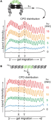
Formation of CPDs in a diverse sequence. (A) The bent 108-mer formed by the 5′-[32P]-108-mer–λ repressor complex (13 pmol) and (B) the linear 108-mer were irradiated at 254 nm in rCutsmart buffer (80 μl, 4°C). Aliquots (1.6 pmol) were removed at the indicated times and digested with T4 polymerase (3 U, 37°C) for 30 min. The resulting DNA fragments were separated by denaturing 8% PAGE and detected by phosphorimagery. The densitometric scans are offset for clarity and indicate the relative quantities of CPDs formed in the 3′-region of the diverse sequence of DNA (highlighted in the helix, see also Supplementary Figures S2 and S4). Sites designated as X (N23) and Y (N19) of the –N33– region represent the two extremes in conformation-dependent CPD formation. The λ repressor is illustrated by the black oval.
Figure 3.
Figure 3.
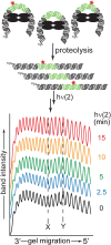
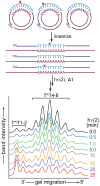
Response of the CPD distribution to irradiation after linearizing a bent DNA. The 5′-[32P]-108-mer–λ repressor complex (27 pmol) was first irradiated ((1)) at 254 nm in rCutsmart buffer (165 μl, 4°C) for 15 min. An aliquot of this bent 108-mer (12 pmol) was then incubated with chymotrypsin (5.5 pmol) at 25°C for 15 min to linearize the DNA. This sample was next irradiated again under identical conditions ((2)). Aliquots (1.6 pmol) were removed at the indicated times of this second irradiation and digested with T4 polymerase (3 U) at 37°C for 30 min. The resulting DNA fragments were separated by denaturing 8% PAGE and detected by phosphorimagery. The 3′-region of the densitometric scans is illustrated here and used to monitor the relative quantities of CPDs (see also Supplementary Figure S8 for full scans). Scans are offset from their origin for clarity and sites defined in Figure 2 are designated as X and Y. The central region of the DNA duplex contains the region of diverse sequence. Stars represent the presence of CPDs and the λ repressor is illustrated by the black oval.
Figure 4.
Figure 4.
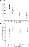
Relative change in CPDs at sites X and Y during a second irradiation with and without prior linearization of bent DNA. Sites X and Y are defined and illustrated in Figures 2 and 3. Aliquots are removed at the indicated times after the second irradiation and processed as described in Figure 3. The difference of peak heights of X and Y prior to the second irradiation is set at 100% and the relative change of this difference is monitored over time of the second irradiation ((2), 254 nm, 4°C) for (A) the bent 108-mer DNA that had been previously irradiated ((1)) and linearized (see Figure 3) and (B) an equivalent sample of bent 108-mer that was previously irradiated ((1)) but not linearized (Supplementary Figure S9). The scatter represents data from six independent experiments. The midline represents their average and the gray box indicates the standard deviation. The dotted line is included to indicate when no difference exists between CPD yields at X and Y.
Figure 5.
Figure 5.
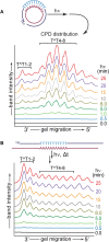
Formation of T^T in T11 DNA. (A) 5′-[32P] Circular T11 DNA (1 pmol) and (B) 5′-[32P] linear T11 DNA (1 pmol) in rCutsmart buffer (130 μl) were irradiated (254 nm) at 4°C. Aliquots (56 fmol) were removed at the indicated times and digested with T4 endonuclease V (10 U, 37°C) for 45 min. Parent DNA and its fragments generated by T4 endonuclease V at sites of T^T were separated with denaturing 10% PAGE and detected by phosphorimagery (see Supplementary Figures S10B and S12B). The densitometric scans are offset for clarity and represent the relative quantities of T^Ts formed at each site within the T11 tract.
Figure 6.
Figure 6.
Response of T^T distribution to linearizing and further irradiating DNA. 5′-[32P] Circular T11 (2 pmol) in rCutsmart buffer (240 μl) was irradiated ((1), 254 nm, 4°C) for 20 min and then an aliquot (1 pmol, 120 μl) was digested with BamHI-HF (100 U, 37°C) for 45 min. The resulting linearized DNA (1 pmol, 130 μl) was further irradiated for 0–25 min at 4°C ((2)). Aliquots (56 fmol) were removed at the indicated times and digested with T4 endonuclease V (10 U, 37°C) for 45 min. Stars represent the presence of T^T. The fragments generated at sites of T^T were separated with denaturing 10% PAGE and detected by phosphorimagery (see Supplementary Figure S14B). The densiometric scans are offset for clarity and indicate the relative quantities of T^T formed at sites within the T11 tract.
Figure 7.
Figure 7.
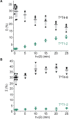
Evolution of the T^T profile after linearizing circular DNA. The shift in T^T distribution from the data illustrated in Figure 6 and Supplementary Figures S14B and S15B is represented for the sum of 3′-sites (T^T1 and T^T2) and the central sites (T^T4–8) after irradiation ((2)) of (A) circular T11 with prior irradiation ((1)) followed by linearization and (B) circular T11 with prior irradiation ((1)) and no linearization. Yields of T^T at each site were calculated by integrating the densitometric scan from valley to valley relative to the total signal of T^T1 through T^T10 and either the linearized T11 (68-mer; see Supplementary Figure S14) for (A) or the circular T11 (80-mer; see Supplementary Figure S15) for (B). The scatter represents data from three independent experiments. The midline represents their average and the gray box indicates the standard deviation.

References

    1. Pfeifer G.P., Besaratinia A.. UV wavelength-dependent DNA damage and human non-melanoma and melanoma skin cancer. Photochem. Photobiol. Sci. 2012; 11:90–97. - PMC - PubMed
    1. Brash D.E. UV signature mutations. Photochem. Photobiol. 2015; 91:15–26. - PMC - PubMed
    1. Jin S.-G., Pettinga D., Johnson J., Li P., Pfeifer G.P.. The major mechanism of melanoma mutations is based on deamination of cytosine in pyrimidine dimers as determined by circle damage sequencing. Sci. Adv. 2021; 7:eabi6508. - PMC - PubMed
    1. Wang S.Y. Photochemistry and Photobiology of Nucleic Acids. 1976; NY: Academic Press.
    1. Mao P., Smerdon M.J., Roberts S.A., Wyrick J.J.. Chromosomal landscape of UV damage formation and repair at single-nucleotide resolution. Proc. Natl Acad. Sci. U.S.A. 2016; 113:9057–9062. - PMC - PubMed

Publication types